
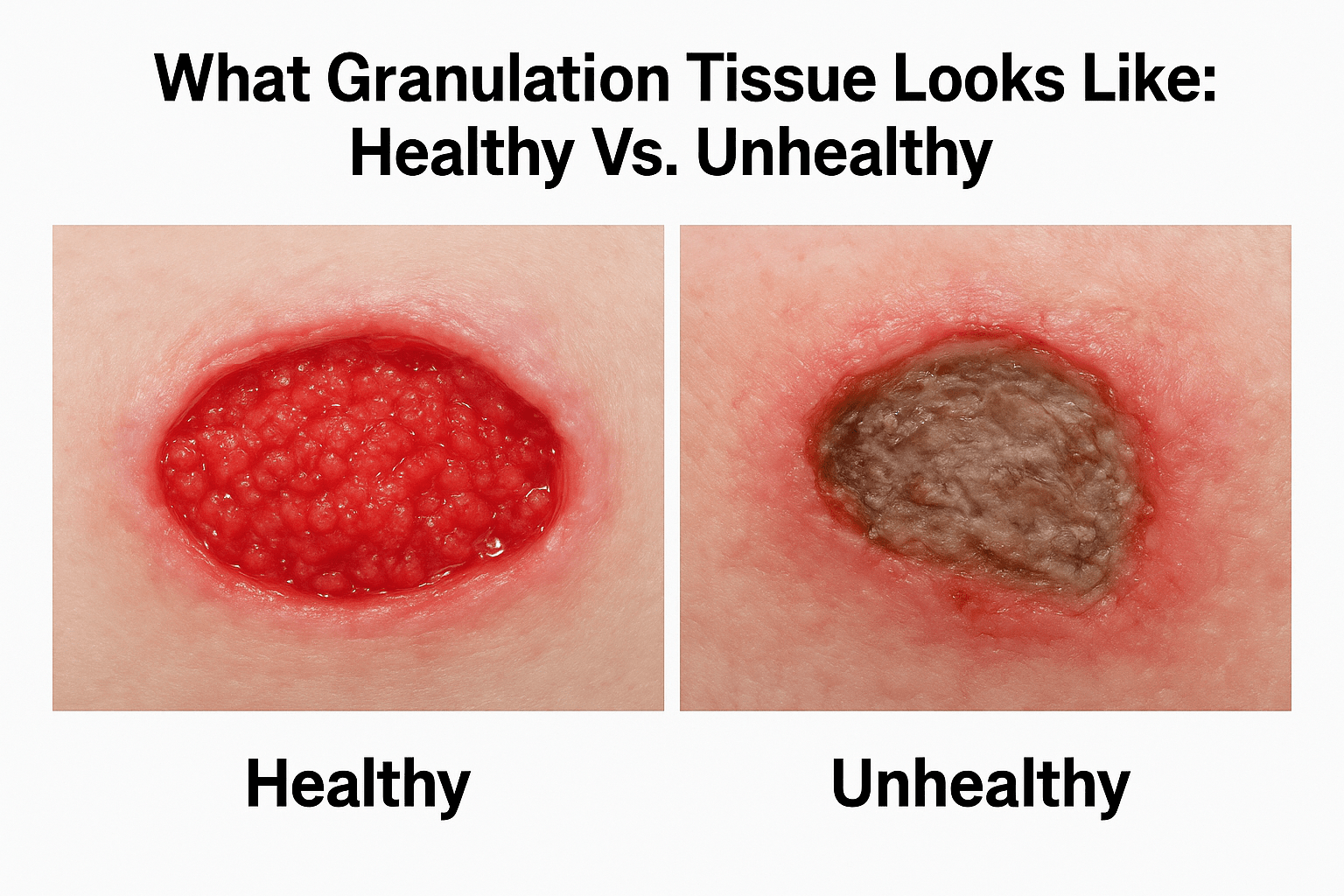
What Granulation Tissue Looks Like: Healthy Vs. Unhealthy
When you’re monitoring a wound, whether on yourself or someone you care for, knowing what granulation tissue looks like can tell you a lot about how recovery is progressing. This beefy, bumpy tissue that fills in an open wound is one of the body’s most visible signs of repair. But not all granulation tissue signals good news. Color, texture, and moisture levels each carry specific meaning, and misreading them can lead to missed complications or unnecessary worry.
At Philadelphia Wound Care, our physician-led mobile practice treats chronic and complex wounds across the Philadelphia region, right at the patient’s bedside. Our General Surgeon evaluates granulation tissue daily in homes, skilled nursing facilities, and hospice settings, so we see the full spectrum of healthy and unhealthy healing firsthand. That clinical experience shapes everything in this guide.
This article breaks down the visual characteristics of granulation tissue at each stage, explains how to distinguish normal healing from warning signs like hypergranulation or pale, friable tissue, and outlines when it’s time to involve a wound care specialist. If you’ve been searching for a clear, reliable reference to assess a wound’s progress, you’re in the right place.
Why granulation tissue appearance matters
Granulation tissue is not just a passive filler. It is an active, living structure that builds the foundation your wound needs to close. When you look at a wound bed and assess what granulation tissue looks like, you are reading a real-time report on how well the body is managing blood supply, infection control, and tissue regeneration. Misreading those signals, or skipping the assessment entirely, can delay treatment decisions by days or even weeks and allow manageable complications to become serious ones.
Getting the visual assessment right early means catching complications before they require more aggressive intervention.
What granulation tissue actually does during healing
The term "granulation" comes from the small, grain-like bumps the tissue forms as new blood vessels grow into the wound bed. Fibroblasts, the cells responsible for producing collagen, migrate into the wound and begin laying down a structural scaffold. Simultaneously, new capillaries extend into this scaffold, giving the tissue its characteristic red or pink color and creating the moist environment that supports cell migration across the wound surface.
This process takes place during the proliferative phase of wound healing, which typically follows the initial inflammatory response. During proliferation, the wound bed should be steadily filling from the bottom up. If you monitor a wound every day or every few days, you will actually see the tissue level rising. That visible upward progress is one of the clearest signs that healing is on track, and losing that progress is one of the earliest signs that something has gone wrong.
How appearance connects to treatment decisions
A wound care provider does not look at a wound simply to confirm it is healing. Every visual characteristic informs a clinical decision, from whether to change the dressing type, to whether a patient needs a tissue culture or a biopsy. Color shifts from bright red to pale pink, gray, or brown are not cosmetic changes. They signal changes in perfusion, moisture balance, or bacterial load, and each one points toward a specific adjustment in the care plan.
Texture matters just as much as color. Tissue that looks raised, friable, or spongy beyond the wound edges may indicate hypergranulation, a condition where the tissue overshoots the wound surface and actually blocks re-epithelialization. Tissue that looks dry, shiny, or stringy may indicate biofilm or inadequate moisture at the wound bed. Both require different management approaches, and neither will resolve on its own without targeted treatment from a qualified provider.
The stages where visual cues carry the most weight
Not every wound stage requires the same level of visual scrutiny. Early-stage wounds, where granulation tissue is just beginning to form, benefit most from close observation because this is when infection or poor perfusion first becomes detectable through visual changes. A wound that stalls at this stage and fails to generate visible granulation within a reasonable timeframe is sending a clear signal that something is disrupting the healing environment, whether that is uncontrolled blood sugar, pressure on the wound site, or an emerging infection.
Later in the healing process, when the wound has mostly filled and epithelialization is beginning at the edges, the appearance of the granulation tissue bed still matters because it directly affects how well new skin cells can migrate across the surface. Dry, uneven, or overgrown tissue creates a physical barrier to final closure. Consistently checking what granulation tissue looks like at this stage helps you and your provider determine whether the wound is truly progressing toward closure or holding at a plateau that requires intervention.
Building this habit of visual observation is not about becoming a medical expert overnight. It is about knowing which changes are worth noting and which are normal so that your care team always has accurate, timely information to act on.
What healthy granulation tissue looks like
When a wound is healing correctly, what granulation tissue looks like follows a consistent pattern you can learn to recognize with practice. The tissue fills in the wound bed steadily, rising from the base upward until it reaches the level of the surrounding skin. Once you understand the specific visual and tactile markers of healthy tissue, you can monitor a wound with much more confidence between clinical visits and give your care team accurate information when you report in.
Color and surface texture
Healthy granulation tissue is bright red to beefy red in color, sometimes compared to the color of raw beef. That vivid color comes from the dense network of new capillaries growing through the tissue to deliver oxygen and nutrients to the wound bed. Small, evenly distributed bumps cover the surface, which is what gives granulation tissue its grainy appearance and where the name originally comes from.
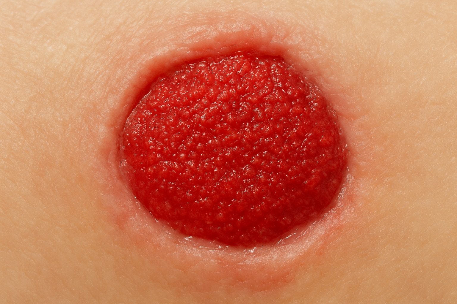
If the tissue in your wound bed matches this description, the wound is in active, productive healing.
The surface should look moist and slightly shiny, not dry or dull. That shine reflects the presence of wound exudate, which keeps the environment hospitable for the fibroblasts and epithelial cells working to close the wound. Uniformity across the wound bed is also a positive sign. Patchy or uneven granulation, where some areas look actively red and healthy while others appear flat or pale, warrants closer attention from your provider.
Moisture and firmness
Healthy granulation tissue holds its shape without being rigid. When gently assessed, it should feel firm but not hard, and it should not bleed easily from light contact. Some minor bleeding during a dressing change is normal given the high capillary density, but tissue that bleeds heavily at the slightest touch is not what you want to see.
The amount of moisture at the wound bed falls within a specific range in a healthy wound. Too little moisture causes the tissue surface to dry out, which slows cell migration and can lead to surface crusting. Too much fluid, particularly if it appears thick or discolored, may signal a shift in the healing environment. Healthy wound exudate is clear to pale yellow and thin, and the skin surrounding the wound should show no signs of maceration or soft tissue breakdown from prolonged moisture exposure.
What unhealthy granulation tissue can look like
Recognizing unhealthy granulation tissue is just as important as spotting healthy healing. When you know what granulation tissue looks like at its worst, you can flag problems quickly and give your care provider the information needed to act before a wound deteriorates further. Three specific patterns show up repeatedly in clinical practice, and each one points to a different underlying problem.
Hypergranulation: tissue that grows too much
Hypergranulation occurs when granulation tissue overfills the wound bed and rises above the surrounding skin level. Instead of stopping at the wound edges, the tissue keeps growing and creates a raised, spongy mound that blocks epithelial cells from migrating across the wound surface. This is one of the most common reasons a wound stalls near the end of the healing process.
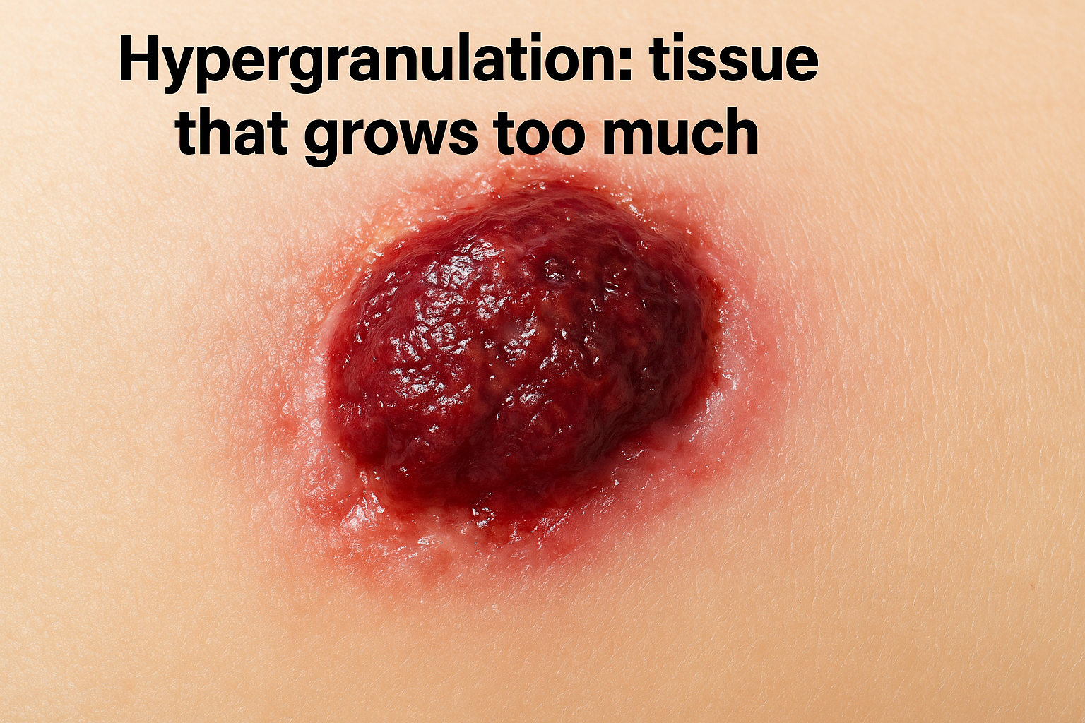
The tissue in a hypergranulating wound often looks dark red, purple-red, or almost burgundy and feels softer and more spongy than healthy granulation. It may bleed easily during dressing changes and appears disproportionately raised compared to the wound margins. Your provider will typically address this by adjusting the dressing type to reduce moisture, applying mild compression, or in some cases using a silver nitrate application to reduce the overgrowth.
Pale, gray, or necrotic tissue
When granulation tissue shifts from bright red to pale pink, gray, yellow, or brown, it signals a drop in blood supply or a significant increase in bacterial load at the wound bed. Pale tissue means fewer functional capillaries are reaching the wound, which directly slows collagen production and cell migration. Gray or brown tissue may indicate early necrosis, where tissue cells are dying from inadequate perfusion or infection.
A wound bed that has shifted to gray or yellow without any area of healthy red granulation needs clinical evaluation promptly, not watchful waiting.
Friable tissue and visible biofilm
Friable granulation tissue bleeds at the lightest touch and falls apart rather than holding its shape. This fragility often signals that a bacterial biofilm has established itself in the wound. Biofilm is a structured colony of bacteria embedded in a protective matrix, and it is one of the main reasons wounds fail to progress despite consistent dressing changes.
You may also notice a dull, slick, or slightly slimy surface on the wound bed alongside friable tissue. That appearance is a strong visual indicator of biofilm rather than normal wound exudate. Biofilm requires mechanical debridement and targeted antimicrobial therapy to disrupt, and standard dressings alone will not resolve it.
How to check and track granulation at home
You do not need clinical training to perform a useful wound assessment at home. Consistent, structured observation between clinical visits gives your care provider an accurate picture of how healing is progressing, and it gives you an early warning system when something changes unexpectedly. The key is knowing exactly what to look for and how to record it so your observations are clear and actionable rather than vague impressions.
What to look for during a dressing change
Dressing changes are your best opportunity to assess what granulation tissue looks like in real time. Before applying the new dressing, take 30 to 60 seconds to examine the wound bed directly under good lighting, ideally natural light or a bright overhead source. Note the color of the tissue: bright beefy red indicates active, healthy healing, while pale pink, gray, or dark purple signals a change worth documenting. Also check whether the tissue sits below the level of the surrounding skin, which means it is still filling in, or whether it has risen above the wound edges, which may indicate hypergranulation.
If the tissue color or height has changed noticeably since the last dressing change, write it down and contact your provider before the next scheduled visit.
Also pay attention to moisture and odor. Healthy wound exudate is thin and clear to pale yellow. Drainage that is thick, cloudy, green, or strongly foul-smelling alongside any color change in the tissue is a combination that warrants prompt clinical contact, not a wait-and-see approach.
Keeping a simple wound log
A wound log does not need to be elaborate to be useful. Recording a few consistent data points after each dressing change builds a timeline that helps your wound care provider make faster, more informed decisions during visits and phone check-ins. Use a notebook or a notes app and log the following each time:
- Date and time of the dressing change
- Tissue color: red, pale pink, gray, or other
- Tissue height: below skin level, level with skin, or raised above edges
- Exudate: amount, color, and thickness
- Odor: absent, mild, or strong
- Surrounding skin condition: intact, red, soft, or breaking down
- Pain or discomfort reported during the change
Photographs add another layer of value. A photo taken from the same angle and distance at each dressing change gives your provider a direct side-by-side visual comparison across multiple days without needing to be physically present. Most smartphones handle this well, and the images can be shared quickly through a patient portal or a follow-up call.
When to call a wound care specialist
Home monitoring is a valuable tool, but it has clear limits. Some changes in what granulation tissue looks like go beyond what a dressing adjustment or an extra observation day can address. Knowing the specific signs that require professional evaluation protects the patient from preventable setbacks and prevents delays that allow manageable complications to become serious wound failures.
Signs that require same-day contact
Some visual changes signal that waiting until the next scheduled visit is not appropriate. Contact your wound care provider the same day if you notice any of the following during a dressing change:
- Tissue color has shifted from red to gray, black, or dark purple across a significant portion of the wound bed
- The wound is producing thick, cloudy, green, or brown drainage combined with a strong foul odor
- The surrounding skin looks red, warm, swollen, or streaked, which may indicate spreading infection
- The patient has developed a fever, chills, or increased pain at or around the wound site
- Bleeding from the wound bed does not stop with gentle pressure within a few minutes
Any combination of color change, increased exudate, and fever is a medical situation that requires prompt clinical evaluation, not self-management at home.
These signs do not always mean the wound is failing, but a qualified physician needs to assess them directly rather than letting them sit until a routine appointment.
Situations where scheduled evaluation is the right move
Not every concern requires an urgent call. Some patterns develop gradually and signal that the current care plan needs a clinical review, even without an acute emergency. Schedule a wound care consultation sooner than planned if the wound bed has shown no visible progress in size reduction or tissue fill over 10 to 14 consecutive days. A plateau at this length without explanation often points to an underlying factor, such as poor perfusion, uncontrolled blood glucose, or biofilm, that standard home dressing changes cannot resolve on their own.
Also bring in a specialist if the granulation tissue appears raised above the wound edges for more than a few days, since hypergranulation rarely self-corrects and blocks the epithelialization needed for final closure. If you are uncertain whether what you are seeing is a normal variation or a warning sign, that uncertainty itself is a reason to call. An experienced wound care physician can answer that question in a single visit and give you a clear path forward.
Simple next steps
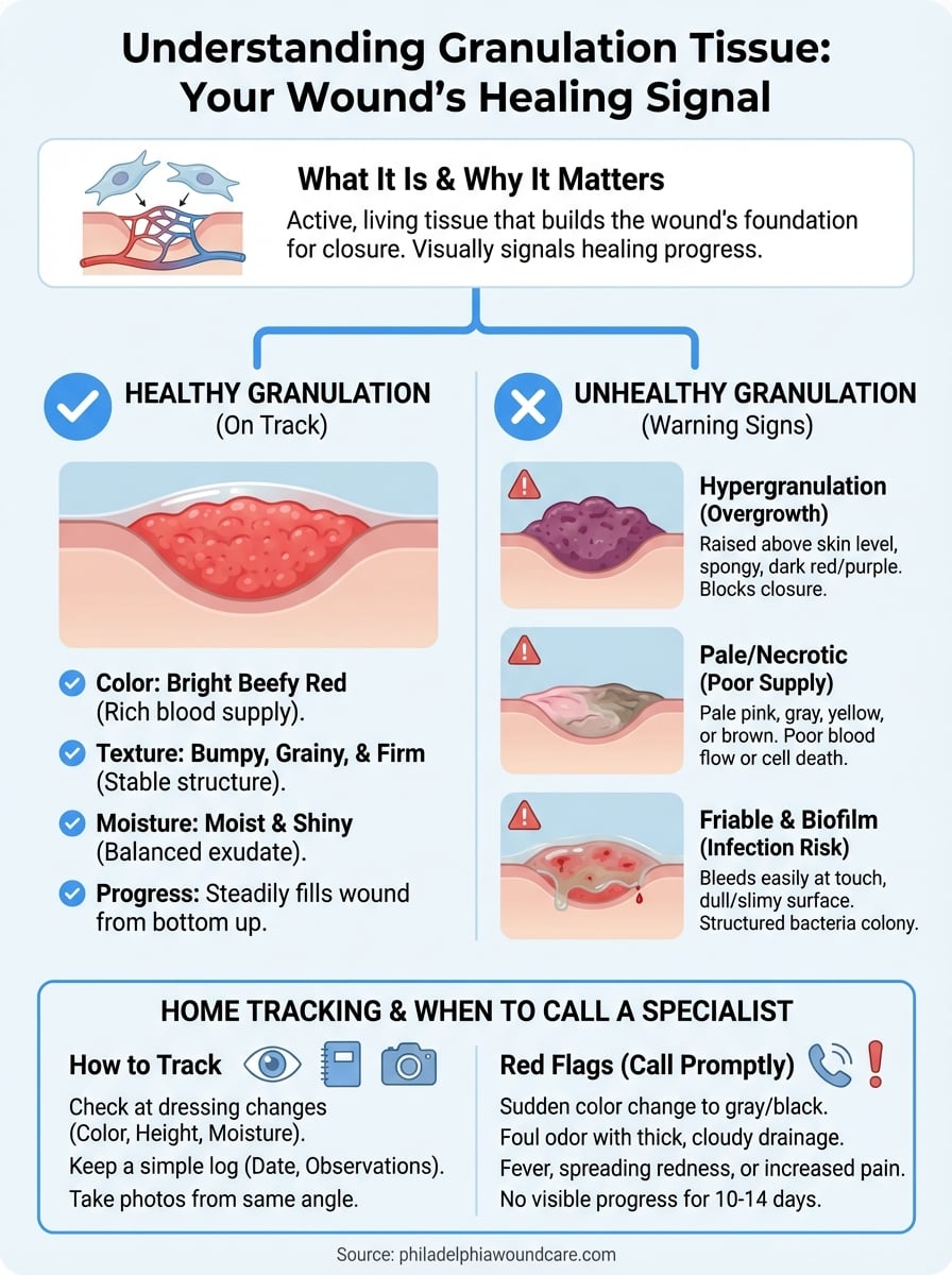
Now you know what granulation tissue looks like when healing is on track and when it signals a problem. Bright red, moist, and evenly filling tissue points to a wound moving in the right direction. Pale, gray, raised, or friable tissue points to something that needs clinical attention before it worsens.
Your most useful next step is building a consistent assessment habit. Check color, moisture, and tissue height at every dressing change, log what you see, and photograph the wound from the same angle each time. That record gives your care team the information they need to adjust your treatment plan quickly and accurately.
If you are in the Philadelphia area and the wound is not progressing as expected, do not wait. A physician-led evaluation at your home or care facility can identify the exact barrier to healing and get treatment started without delay. Reach out to Philadelphia Wound Care’s mobile wound care team to schedule an assessment.