
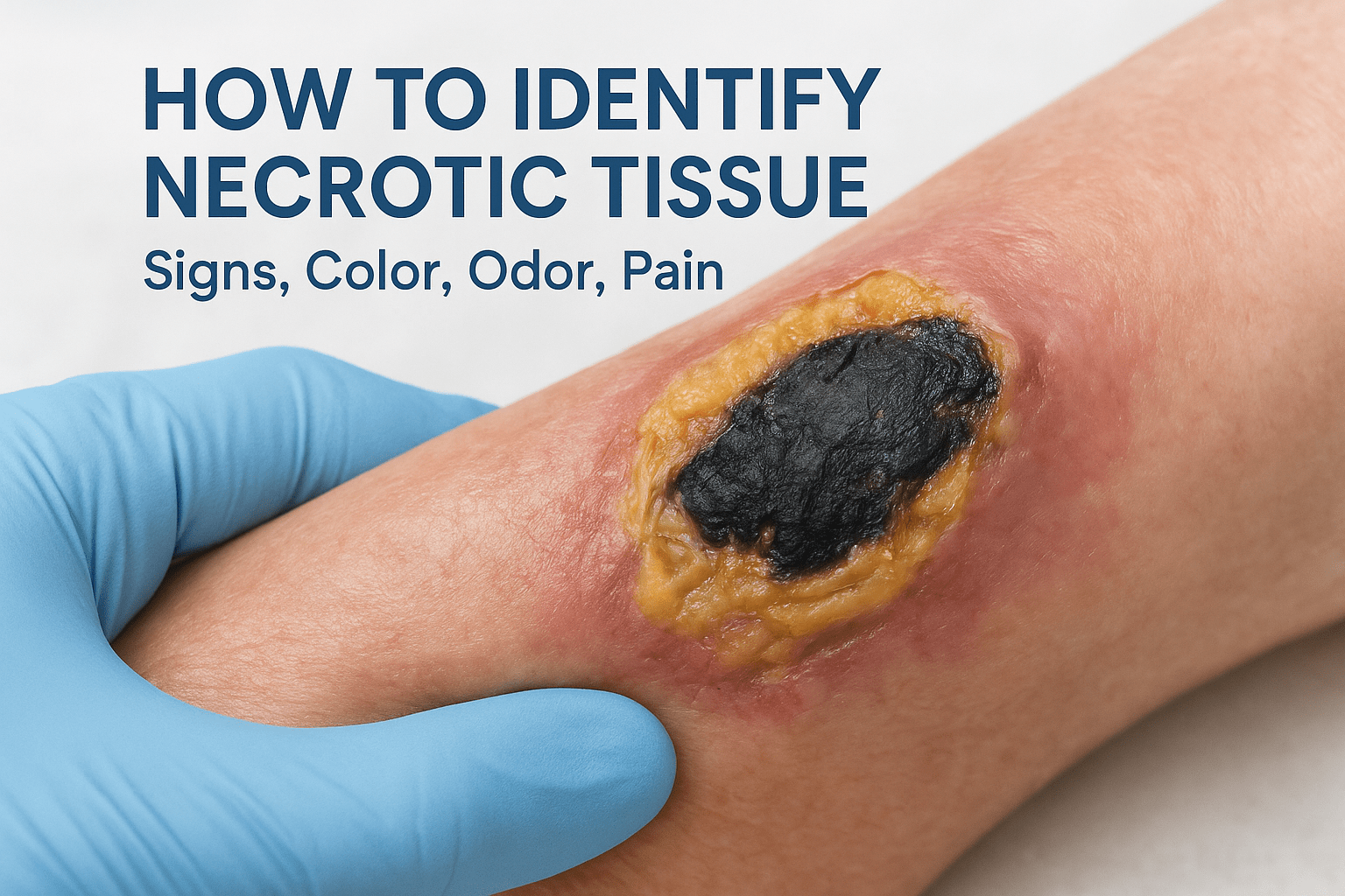
How To Identify Necrotic Tissue: Signs, Color, Odor, Pain
A wound that isn’t healing deserves a closer look, especially when the tissue itself starts to change. Knowing how to identify necrotic tissue is one of the most important skills a caregiver or patient can develop, because dead tissue in a wound bed actively blocks recovery and raises the risk of serious infection. The color, texture, odor, and pain level of wound tissue all tell a story, and reading those signals early can mean the difference between a wound that heals and one that gets worse.
At Philadelphia Wound Care, our physician-led mobile practice treats chronic and complex wounds at the patient’s bedside, in homes, skilled nursing facilities, and hospice settings across the Philadelphia area. We routinely evaluate tissue viability as part of every wound assessment. Necrotic tissue is something we see often, particularly in patients managing diabetic ulcers, pressure injuries, and vascular wounds. It’s also one of the most common reasons families reach out to us: they notice something in the wound that doesn’t look right and want answers.
This article breaks down the specific visual and physical signs of necrotic tissue, including what different colors mean, when odor signals a deeper problem, and how pain patterns can point to tissue death. Whether you’re a caregiver monitoring a loved one’s wound or a facility nurse coordinating next steps, this guide gives you the practical knowledge to recognize necrotic tissue and understand when it’s time to call in a specialist.
Why identifying necrotic tissue matters
Necrotic tissue is dead tissue, and dead tissue in a wound does not stay neutral. It creates an environment where bacteria thrive and infection spreads, pulling the wound further from any possibility of natural recovery. When you understand how to identify necrotic tissue early, you give clinicians the information they need to act before complications compound. Every day of delay matters when tissue death is involved, particularly in patients who already carry risk factors like diabetes, poor circulation, or immunosuppression.
It sets off a chain reaction in the wound
Dead tissue acts as a physical barrier that blocks new skin cells from migrating across the wound surface. Healthy tissue cannot grow into or through a layer of hardened, dried, or liquefying material. The wound stalls, and what started as a manageable injury can escalate into a problem that reaches deep tissue, bone, or the bloodstream. Sepsis, osteomyelitis, and limb amputation are not rare outcomes when necrotic wounds go unrecognized for extended periods, especially in elderly patients or those with compromised immune systems.
Identifying necrotic tissue early is not just about the wound itself. It is about preventing systemic consequences that can become life-threatening.
Patients with diabetic foot ulcers and pressure injuries face some of the highest rates of tissue necrosis, and these are also the populations least likely to feel early warning symptoms because of neuropathy or reduced sensation. That gap between physical damage and perceived pain is one reason caregivers and family members play such a critical role in wound monitoring.
It directly shapes the treatment plan
Not all necrotic tissue presents the same way, and the type present in a wound directly determines which treatment approach a physician will use. Dry, black eschar is managed differently from soft, wet slough. When you bring accurate observations to a specialist, including color changes, texture, and odor, you accelerate the diagnostic process and help the care team choose the right intervention faster.
Consistent monitoring between clinical visits also gives providers more reliable data about how a wound is changing. That information reduces the chance that a dangerous shift in wound condition goes unnoticed until the next scheduled appointment.
Necrotic tissue vs healthy healing tissue
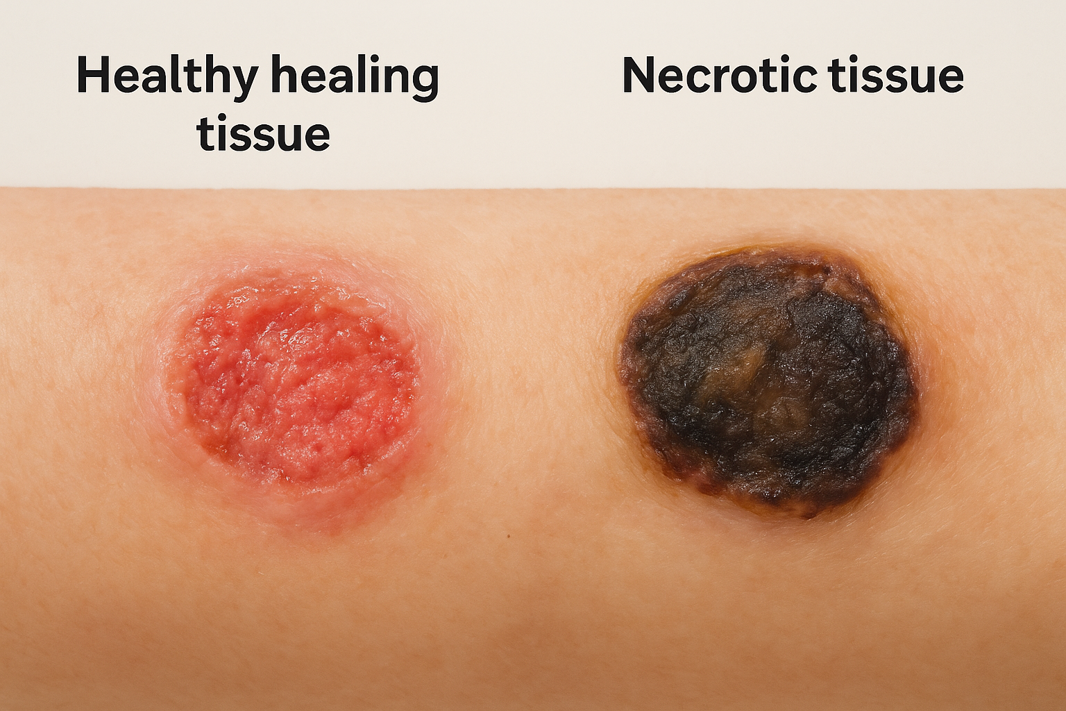
Knowing how to identify necrotic tissue becomes much easier once you understand what healthy, healing tissue actually looks like. A wound that is progressing normally has a distinct set of visual characteristics, and recognizing the contrast between living and dead tissue helps you spot problems before they get worse.
What healthy tissue looks like
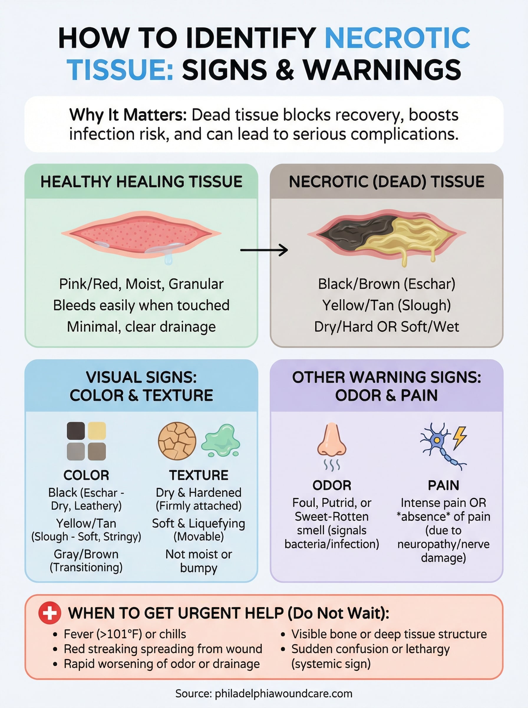
Healthy granulation tissue is pink or red, moist, and slightly bumpy in texture. It bleeds easily when touched, which signals an active blood supply feeding the wound bed. New skin grows inward from the edges, and drainage stays minimal, clear, or faintly yellow. A wound on track looks incrementally better from one visit to the next.
When tissue color stays consistent and pink, and edges actively close, healing is happening. When color shifts or drainage changes character, something in the wound has changed.
What necrotic tissue signals by comparison
Necrotic tissue shares none of those qualities. Instead of a pink, vascular surface, you find tissue that has shifted to yellow, brown, gray, or black. The texture moves away from moist and granular toward either dry and hardened or soft and liquefying, depending on the type of cell death occurring. The wound often stops changing for the better entirely.
Your observation of the wound edges also matters here. Rather than skin visibly migrating inward, you may notice edges that appear flat, thickened, or rolled, which indicates that new cell growth has stalled. Dead tissue physically blocks that migration, and any shift from a healthy wound bed toward discolored or deteriorating tissue warrants immediate contact with a wound care specialist.
Visual signs: color, texture, and drainage
Color is the first thing most caregivers notice when tissue begins to die. Learning how to identify necrotic tissue starts with understanding that color shifts follow a predictable pattern, and each shade carries a specific clinical meaning. What you see in the wound bed gives you and any clinician you contact an immediate, concrete picture of how far tissue death has progressed.
Color: what different shades tell you
Wound tissue color maps directly to the type and stage of necrosis present. Black tissue, called eschar, is typically dry, firm, and leathery, indicating that the tissue has completely lost circulation and hardened over time. Yellow or tan soft tissue, referred to as slough, signals a mix of dead cells, fibrin, and bacteria that has not yet dried out, leaving a looser, stringy material sitting across the wound surface.
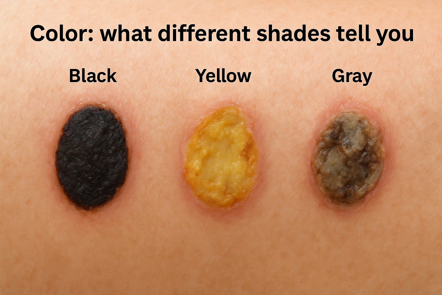
Gray or brown tissue that appears wet or discolored often indicates that the wound is transitioning between stages, which makes early physician evaluation critical.
Texture and drainage changes
Texture gives you as much information as color does. Dry, hardened tissue firmly attached to the wound base almost always points to eschar, while soft, easily movable material that shifts when touched suggests slough. Neither type shares the moist, granular, slightly bumpy quality you would expect from healthy granulation tissue actively rebuilding beneath the surface.
Drainage also changes character when necrosis is present. Rather than minimal clear or faintly yellow fluid, you may notice thick, murky, greenish, or brown discharge accumulating around the wound. A combination of increased drainage volume and visible tissue discoloration is a reliable, observable signal that dead material is present and requires professional assessment.
Smell, pain, and other warning symptoms
Color and texture give you strong visual evidence of tissue death, but odor and pain levels complete the clinical picture when you are learning how to identify necrotic tissue. A wound producing a foul or unusually strong smell communicates something specific: bacterial activity in dead tissue generates distinct byproducts that create an unmistakable odor differing sharply from the mild scent of a normally healing wound.
Odor as a clinical signal
When you notice a wound that smells putrid, sweet-rotten, or particularly sharp, that odor typically indicates bacteria have colonized necrotic tissue and begun breaking it down. Different bacteria produce different smells, which gives clinicians useful diagnostic information:
- Pseudomonas aeruginosa: sweet or fruity odor
- Anaerobic bacteria: fecal-like or strongly offensive odor
- Proteus species: ammonia-like smell
A strong, foul odor from a wound is not something to normalize or manage with dressings alone; it signals that professional evaluation is needed.
Pain patterns and their absence
Pain in a wound can point toward necrosis, but the absence of pain can be equally alarming. Patients with intact sensation may report a burning, throbbing, or deep ache around a deteriorating wound. However, patients with diabetic neuropathy or advanced vascular disease often feel little to nothing even as tissue death progresses, which is why pain level alone should not determine how seriously you take a wound.
Surrounding skin that feels cool, looks dusky, or appears visibly swollen next to the wound also signals that tissue perfusion has decreased. These additional physical signs, paired with odor changes, give you a more complete assessment picture to share with a wound care specialist when you call.
When to get urgent help and what to expect
Once you know how to identify necrotic tissue, the next step is understanding which signs require same-day or emergency action rather than a routine appointment. Necrosis that spreads quickly, or tissue death accompanied by systemic symptoms, moves beyond standard outpatient wound care into urgent medical intervention.
Signs that cannot wait
Some changes in a wound demand immediate contact with a physician or emergency services. If you notice any of the following alongside visible necrotic tissue, do not wait:
- Fever above 101°F or chills
- Red streaking spreading outward from the wound edges
- Rapid worsening of odor or a significant increase in drainage volume
- Sudden confusion or unusual lethargy in the patient
- A wound that visibly reaches bone or joint structures
Systemic symptoms like fever and confusion indicate that infection has moved beyond the wound itself and may be entering the bloodstream, which shifts this from a wound problem to a whole-body emergency.
When a wound shows local signs of necrosis alongside systemic symptoms, that combination signals sepsis risk and requires emergency evaluation without delay.
What a specialist evaluation involves
When you contact a wound care physician, expect a thorough assessment of tissue viability, wound depth, and surrounding skin condition. The clinician will document color, texture, odor, and drainage, then determine whether debridement is needed to remove dead material. Debridement, whether sharp, enzymatic, or biological, clears the wound bed so healthy tissue can finally grow. Your physician may also order imaging or laboratory work to rule out bone infection or systemic spread before finalizing a treatment plan.
What to do next
Knowing how to identify necrotic tissue gives you the practical foundation to act before a wound deteriorates beyond the point where simple interventions work. You now understand that color, texture, odor, and pain patterns each carry distinct clinical information, and that specific combinations of those signs require urgent physician contact rather than a watch-and-wait approach. That knowledge has real consequences for the person whose wound you are monitoring.
Your next step is straightforward: if you have seen any of the signs described in this article, do not manage the situation with home dressings alone. Philadelphia Wound Care brings physician-led mobile wound care directly to patients in homes, skilled nursing facilities, and hospice settings across the Philadelphia region, with a 24-hour response time for urgent needs. Reach out to our team through our mobile wound care services page to request an evaluation or learn more about how we can help.