

Bil Lower Extremity Edema: Causes, Workup, And Treatment
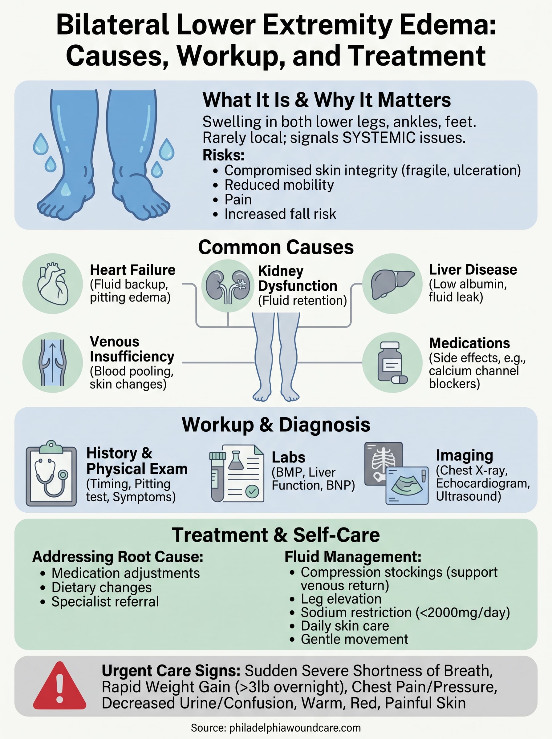
When both legs begin to swell at the same time, it’s rarely a local problem. Bil lower extremity edema, bilateral swelling of the lower legs, ankles, or feet, typically signals something systemic going on in the body. It could point to heart failure, kidney dysfunction, liver disease, or a handful of other conditions that demand prompt evaluation and management.
This type of edema is especially common among older adults and individuals with chronic illnesses like diabetes or vascular disease. Left unaddressed, the persistent swelling doesn’t just cause discomfort, it compromises skin integrity and creates conditions ripe for wound formation. Skin stretched thin by fluid retention breaks down faster, heals slower, and becomes far more vulnerable to ulceration and infection, particularly in the lower legs where circulation is already working against gravity.
At Philadelphia Wound Care, our physician-led mobile practice treats the downstream consequences of bilateral edema on a daily basis. Dr. Weber sees firsthand how unmanaged swelling leads to chronic wounds, venous ulcers, skin breakdown, and surgical sites that refuse to close, in patients across Philadelphia’s homes, skilled nursing facilities, and assisted living communities. That clinical reality is exactly why understanding the causes, workup, and treatment of this condition matters so much.
This article breaks down what bilateral lower extremity edema is, why it develops, how clinicians evaluate it, and what treatment looks like, giving patients, caregivers, and referring providers a clear, practical reference to guide next steps.
Why bilateral leg swelling matters
Bilateral leg swelling is not a cosmetic issue or a minor inconvenience. When you see both legs affected at the same time, it tells you the body is struggling to maintain fluid balance at a systemic level. Unlike unilateral swelling, which often points to something localized like a clot or injury, bilateral edema typically reflects a widespread physiological problem that needs a thorough workup, not watchful waiting.
The skin pays the price
Skin stretched by excess fluid becomes compromised in ways that are easy to underestimate. The outer layers lose their structural integrity as fluid pushes from beneath, making the surface fragile, dry, and prone to small tears. Even modest pressure from a shoe, a sock band, or minor friction against bedsheets can create an opening that, in a healthy person, would heal quickly, but in someone with chronic bilateral edema can evolve into a non-healing wound.
Persistent lower extremity edema is one of the most common underlying factors driving venous leg ulcers, which account for roughly 70% of all chronic leg wounds.
For patients managing diabetes or peripheral vascular disease, this risk compounds significantly. Reduced circulation and compromised nerve function mean the body sends fewer warning signals when skin breaks down. By the time someone notices a wound, it may already be deep, infected, or surrounded by fragile tissue that resists standard healing efforts.
Mobility and independence take a direct hit
Swollen, heavy legs make basic movement painful and exhausting. For older adults, reduced mobility accelerates a cycle of deconditioning that raises fall risk, worsens cardiovascular function, and increases dependence on caregivers. Many patients describe the sensation as carrying extra weight they cannot set aside, which discourages activity and leads to further fluid accumulation from prolonged sitting or lying down.
Reduced activity also affects wound healing directly. Movement promotes venous return, the process by which blood travels back up the legs toward the heart. Without regular calf muscle contraction, fluid pools more aggressively in the lower legs, and tissue oxygenation drops in exactly the areas where your body needs it most to prevent or recover from skin breakdown.
Why recognizing bil lower extremity edema early changes outcomes
Catching bilateral edema early gives you and your care team the best chance of addressing the root cause before complications develop. When systemic conditions like heart failure or kidney disease are identified and managed in earlier stages, the edema often responds well to medication adjustments, dietary changes, and compression therapy. At that point, you are managing a chronic condition rather than repairing an open wound or treating sepsis from an infected ulcer.
Late-stage management is harder, costlier, and harder on the patient. Once skin breakdown has occurred, treatment timelines stretch from days to months. Patients who might have managed at home require hospitalization or intensive outpatient wound care. For frail or elderly patients, a single non-healing ulcer can accelerate overall functional decline in ways that reach well beyond the wound itself.
Common causes and what they suggest
When you see bilateral lower extremity edema, the cause is almost always one of a handful of well-defined systemic conditions. Identifying the underlying driver is not just an academic exercise; it directly shapes your treatment plan and determines how urgently the patient needs evaluation.
Cardiac, renal, and hepatic causes
Heart failure is the most common systemic cause of bilateral leg swelling in older adults. When the heart cannot pump efficiently, fluid backs up into the venous system and settles in the dependent parts of the body, primarily the legs. The swelling tends to be pitting, meaning a fingertip pressed into the skin leaves an indentation, and it typically worsens as the day progresses.
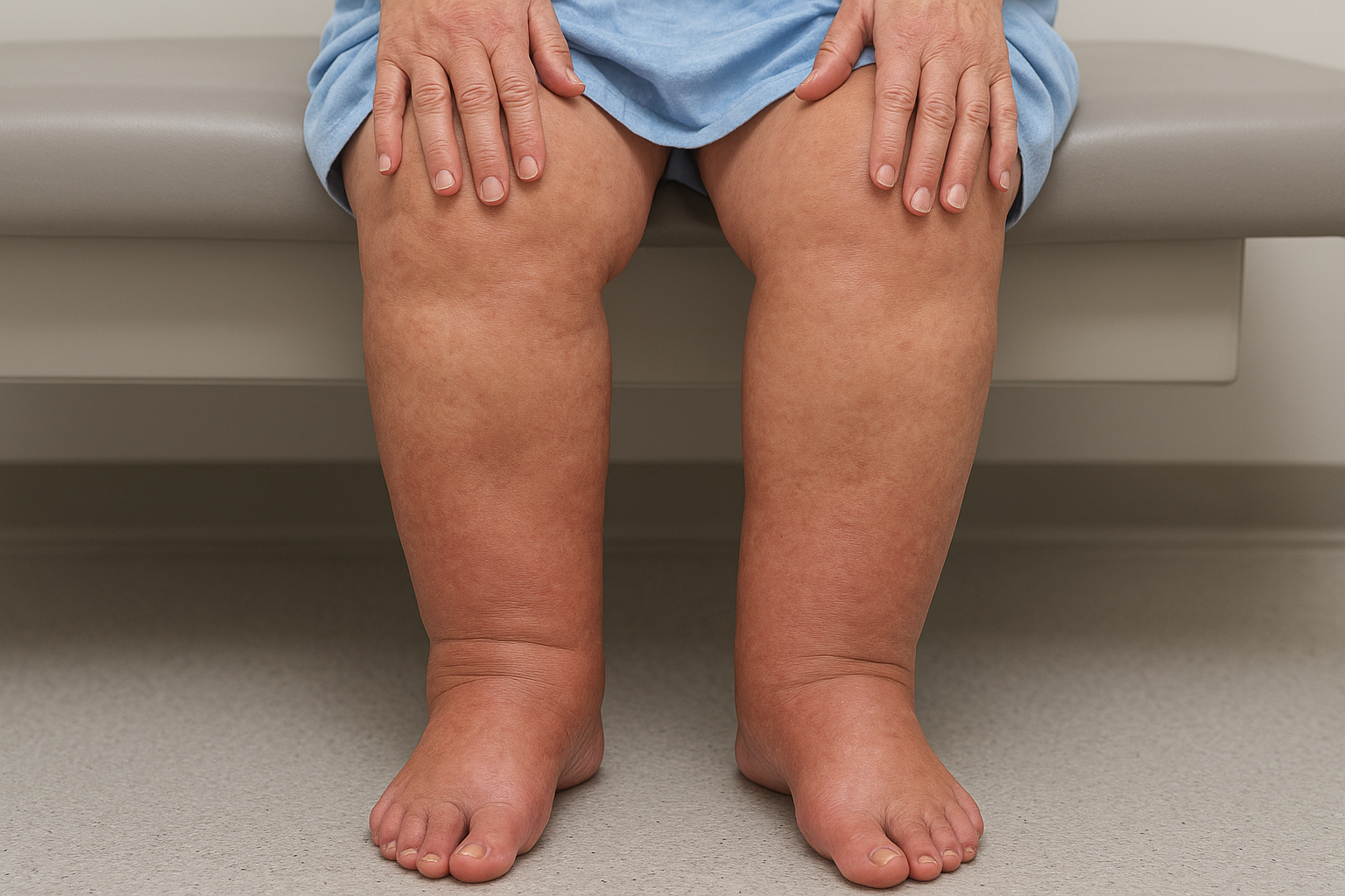
Kidney disease impairs the body’s ability to excrete sodium and water, leading to total-body fluid retention that shows up first in the legs. Hepatic cirrhosis causes bilateral edema through a different mechanism: falling albumin levels reduce oncotic pressure in the bloodstream, allowing fluid to leak into surrounding tissues. Both conditions usually come with additional clinical signs that help distinguish them from cardiac edema.
When bilateral edema accompanies shortness of breath, rapid weight gain, or abdominal distension, these findings together point strongly toward a cardiac, renal, or hepatic origin and warrant immediate workup.
Venous insufficiency and medication effects
Chronic venous insufficiency is another major driver of bil lower extremity edema, particularly in patients who spend long hours sitting or standing. Incompetent venous valves allow blood to pool in the lower legs, creating sustained pressure that forces fluid into surrounding tissue. Over time, this leads to skin changes like hyperpigmentation, lipodermatosclerosis, and eventually ulceration, which is exactly where wound care intervention becomes necessary.
Certain medications also produce bilateral swelling as a direct side effect. Calcium channel blockers, commonly prescribed for hypertension, are among the most frequent culprits. Corticosteroids, NSAIDs, and some diabetes medications carry the same risk. Reviewing a patient’s full medication list is a straightforward step that can sometimes explain edema appearing resistant to other treatments, without requiring extensive additional testing.
How clinicians work up bilateral leg edema
When a patient presents with bilateral leg swelling, a clinician’s first job is to narrow down the cause systematically. The workup for bil lower extremity edema follows a logical sequence: history, physical examination, targeted labs, and imaging when indicated. Jumping straight to testing without a structured clinical approach wastes time and often leads to a missed or delayed diagnosis.
Starting with the history and physical exam
A focused history tells you more than most tests will. You want to know how long the swelling has been present and whether it worsens throughout the day, then partially resolves overnight with leg elevation. Asking about associated symptoms such as shortness of breath, rapid weight gain, abdominal fullness, or decreased urine output points you toward cardiac, hepatic, or renal causes almost immediately.
The physical exam adds critical detail. Pressing firmly on the lower leg or ankle for a few seconds tells you whether the edema is pitting or non-pitting, which helps distinguish fluid overload from lymphedema or lipedema. Checking for jugular venous distension, lung crackles, or an enlarged liver during the same visit can confirm a systemic origin without any lab results in hand.
Lab work and imaging
Once your history and exam have shaped your suspicion, targeted lab work confirms it. A basic metabolic panel tells you about kidney function and electrolyte balance, while liver function tests and albumin levels identify hepatic involvement. A brain natriuretic peptide (BNP) or NT-proBNP level is the most sensitive initial test for heart failure and should be ordered whenever cardiac edema is on the differential.
An elevated BNP combined with bilateral leg swelling and exertional shortness of breath gives you a strong presumptive diagnosis of heart failure before any imaging is done.
Imaging comes into play when the diagnosis remains unclear or when you need to quantify the underlying problem. A chest X-ray can reveal pulmonary congestion or pleural effusions consistent with cardiac failure. An echocardiogram gives you a direct look at cardiac function and is the standard next step when heart failure is suspected. Doppler ultrasound of the lower extremities is less commonly ordered for bilateral edema but becomes relevant when you cannot confidently rule out simultaneous deep vein thrombosis in both legs.
Treatment options and self-care steps
Treating bil lower extremity edema effectively requires two parallel tracks: managing the root cause and directly controlling the fluid accumulation. If you only address the swelling without identifying why it developed, the edema returns as soon as the intervention stops. Working through both tracks simultaneously gives patients the best chance of reducing swelling, protecting skin integrity, and preventing wound formation.
Addressing the underlying cause
The condition driving the edema determines first-line medical treatment. Heart failure is typically managed with diuretics, ACE inhibitors or ARBs, and beta-blockers, alongside strict sodium restriction and daily weight monitoring. Kidney disease may require fluid intake adjustments, nephrology referral, or dialysis if function has deteriorated significantly. For hepatic causes, the goal is managing cirrhosis and its complications, including albumin replacement when levels drop critically low.
When a medication review identifies a drug-induced cause, such as a calcium channel blocker, coordinating with the prescribing physician to switch or adjust the dose can resolve the edema without adding new treatments.
A structured review of the full medication list should happen early in every workup. Corticosteroids, NSAIDs, and certain diabetes medications all carry edema as a recognized side effect, and catching this early can spare patients unnecessary testing or treatment escalation.
Compression therapy and leg elevation
Compression stockings remain one of the most effective non-pharmacological tools for bilateral lower extremity swelling, particularly when venous insufficiency is a contributing factor. Graduated compression between 20 and 40 mmHg applies consistent external pressure that supports venous return and reduces fluid pooling. Patients need proper fitting and instruction on application; an ill-fitted stocking can cause pressure injuries in patients with fragile or already-compromised skin.
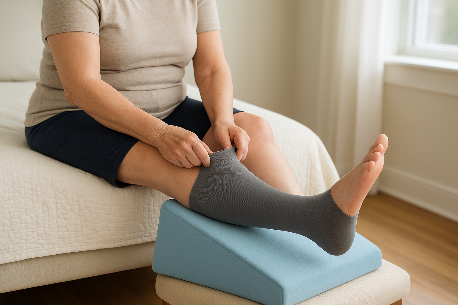
Encouraging leg elevation above heart level for 30-minute intervals throughout the day works alongside compression by using gravity to move fluid back toward central circulation. For bedbound or mobility-limited patients, positioning wedges placed under the mattress achieve the same effect passively and consistently.
Daily self-care habits that support treatment
Patients and caregivers can take concrete daily steps that reinforce medical treatment. Key habits include:
- Sodium restriction to under 2,000 mg per day to limit fluid retention
- Daily skin inspection of the lower legs to catch early breakdown before it becomes a wound
- Gentle moisturizing of dry or stretched skin to reduce cracking and fissure formation
- Light walking when mobility allows, to activate calf muscles and promote venous return
When to seek urgent care and when to refer
Not every case of bil lower extremity edema demands an emergency room visit, but some presentations signal a medical crisis that cannot wait for a scheduled appointment. Knowing the difference between urgent and routine helps patients, caregivers, and referring providers act at the right speed without either overcrowding emergency departments or delaying care that could prevent serious harm.
Signs that require emergency evaluation
Some symptoms appearing alongside bilateral leg swelling tell you that something dangerous is happening right now. These are not situations where you monitor the patient at home and see how they feel in the morning. They require same-day emergency evaluation, often by calling 911 or going directly to an emergency department.
Seek urgent care immediately if bilateral lower extremity edema accompanies any of the following:
- Sudden, severe shortness of breath or inability to lie flat without feeling breathless
- Rapid weight gain of more than three pounds overnight or five pounds in a week
- Chest pain or pressure, which could indicate decompensated heart failure or a cardiac event
- Significantly decreased urine output combined with confusion or lethargy, suggesting acute kidney failure
- Skin that is warm, red, and painful in both legs, raising concern for bilateral cellulitis
When bilateral leg swelling comes on suddenly rather than developing gradually over weeks, the pace of onset itself is a red flag that warrants urgent evaluation regardless of other symptoms.
When to refer to a specialist
Outside of emergency situations, certain clinical patterns signal that primary care management alone will not be sufficient to resolve the edema or protect the patient from complications. If your initial workup points toward advanced heart failure, cirrhosis with ascites, or nephrotic syndrome, connecting the patient with the relevant specialist, a cardiologist, hepatologist, or nephrologist, ensures the root cause receives the level of attention it requires.
Referral to a wound care specialist becomes the right call when bilateral edema has already contributed to skin breakdown. Venous ulcers, pressure injuries, and post-surgical wounds that form in the setting of chronic swelling heal poorly without targeted intervention. A physician-led mobile wound care service can reach patients who cannot travel to a clinic, bringing advanced therapies directly to the patient’s bedside and closing the gap between hospital discharge and full recovery.
Next steps
Bilateral lower extremity edema sits at the intersection of systemic disease and skin integrity. Understanding the underlying cause, working through a structured clinical evaluation, and applying the right combination of medical treatment and compression therapy gives you the best chance of controlling the swelling before it turns into a chronic wound. Early identification of the root condition and a coordinated care approach consistently produce better outcomes than waiting until skin breakdown forces a more intensive intervention.
If you or someone you care for is managing bil lower extremity edema that has already led to skin breakdown, venous ulcers, or wounds that are not progressing, getting physician-led wound care to the patient’s location removes the barrier of travel entirely. Request mobile wound care services in Philadelphia to connect with a specialist who can evaluate and treat complex wounds directly at the bedside, wherever the patient is.