

Stages Of Biofilm Development: Attachment To Dispersion
Biofilms are one of the most persistent obstacles to wound healing. These structured communities of bacteria anchor themselves to wound beds, shield themselves from antibiotics, and actively resist the body’s immune response. For patients managing chronic or non-healing wounds, diabetic ulcers, pressure injuries, venous ulcers, biofilm presence is often the reason a wound stalls and refuses to close. Understanding the stages of biofilm development gives both clinicians and patients a clearer picture of what’s happening at the wound surface and why timely intervention matters.
A biofilm doesn’t appear overnight as a fully formed barrier. It builds through a specific biological sequence, starting with a single bacterium attaching to a surface and progressing through growth, maturation, and eventual dispersal to new sites. Each stage presents distinct characteristics and treatment implications, and recognizing where a biofilm sits in that progression directly informs clinical decision-making.
At Philadelphia Wound Care, our physician-led mobile practice treats complex wounds at the bedside, in homes, skilled nursing facilities, and assisted living communities across the Philadelphia area. Biofilm management is a routine part of our clinical work, particularly when wounds have resisted standard care. This article breaks down each stage of biofilm development from initial attachment through dispersion, explaining the biology behind the process and why it matters for anyone dealing with a wound that won’t heal.
What biofilms are and how they form
A biofilm is a structured community of microorganisms, most commonly bacteria, that attach to a surface and encase themselves in a self-produced protective matrix. This matrix, made of polysaccharides, proteins, and nucleic acids, is called the extracellular polymeric substance, or EPS. It functions as a biological shield the bacteria manufacture themselves. Inside that shield, bacteria behave very differently than they do in their free-floating, or planktonic, form. They communicate, share resources, and collectively resist threats that would kill individual cells with ease.
The biology behind biofilm structure
Biofilms are not a random accumulation of bacteria stuck together. They are highly organized communities with distinct internal architecture. Within a mature biofilm, you will find water channels that distribute nutrients and remove waste, functioning much like a primitive circulatory system. Different bacterial species can co-exist in a single biofilm, and genetic exchange between those species occurs far more readily inside the biofilm than in planktonic environments. This genetic mixing can accelerate the development of antibiotic resistance, making treatment significantly more difficult as time passes.

The EPS matrix surrounding a biofilm community can account for up to 90% of the biofilm’s total mass, with the bacteria themselves making up a relatively small fraction of the overall structure.
Biofilm structure also allows these communities to form on a wide range of surfaces, from medical implants and catheters to the tissue of a chronic wound. On wound beds, bacteria exploit damaged, poorly vascularized tissue that the immune system struggles to patrol effectively. That tissue gives the biofilm a stable platform to anchor and expand.
How bacteria build a biofilm from scratch
To understand the stages of biofilm development, you first need to understand where the process begins. Free-floating bacteria encounter a surface, whether it is a wound bed, necrotic tissue, or a foreign body, and begin evaluating whether local conditions support attachment. This initial contact is driven by physical and chemical signals in the surrounding environment, including moisture levels, nutrient availability, and surface properties like texture and electrical charge.
Once bacteria detect a favorable surface, they begin to change at a genetic level. They alter gene expression, shifting from planktonic mode to surface-attached mode. Flagella, the tail-like appendages bacteria use to propel themselves through fluid, are downregulated. Instead, bacteria start producing the molecular adhesins and early EPS components that allow them to stick. At this point, the attachment is still reversible. A sufficient physical disruption or shift in conditions can dislodge these early surface settlers.
What follows that first contact is a cascade of biological events, each stage building directly on the last, that transforms a small cluster of surface-attached bacteria into a fully mature, antibiotic-tolerant biofilm. Proteins on the bacterial surface bind to host tissue proteins like fibronectin and collagen, anchoring the colony more firmly. Quorum sensing, the chemical communication system bacteria use to coordinate collective behavior, becomes increasingly active as cell density rises. Each of these steps is driven by survival pressure, and each one makes the biofilm harder to disrupt than the step before it.
Why biofilms matter in wound care
Biofilms are estimated to be present in 60 to 80 percent of chronic wounds, making them one of the most common and clinically significant barriers your wound care team will face. When bacteria form a biofilm on a wound bed, they stop behaving as individual organisms and start functioning as a coordinated, defensive community. That shift fundamentally changes how difficult the infection is to treat and how long your wound will take to heal.
The clinical impact on chronic wounds
A wound that contains a biofilm does not simply heal more slowly. It often stops progressing entirely. The biofilm disrupts the normal healing cascade by triggering a persistent, low-grade inflammatory response that prevents the tissue remodeling your body needs to close the wound. Instead of moving through the stages of inflammation, proliferation, and remodeling, the wound stays locked in a chronic inflammatory state. Clinicians often describe this as a wound that appears "stuck" without a clear reason, presenting with stalled granulation tissue and ongoing drainage despite consistent care.
Biofilm-associated wounds can appear deceptively clean on the surface, showing minimal redness or visible infection, while the bacterial community underneath continues driving tissue damage and blocking healing.
For patients managing diabetic foot ulcers, pressure injuries, or venous leg ulcers, this stalling effect is particularly dangerous. Prolonged inflammation contributes to further tissue breakdown, and a wound that won’t close raises the risk of systemic infection, osteomyelitis, and in severe cases, amputation.
Why standard antibiotics fall short
Understanding the stages of biofilm development helps explain exactly why oral and topical antibiotics frequently fail to resolve biofilm-related wound infections. Once bacteria have established their EPS matrix and transitioned into biofilm mode, antibiotic concentrations that would eliminate planktonic bacteria become almost entirely ineffective. Research has shown that bacteria inside a mature biofilm can tolerate antibiotic concentrations 100 to 1,000 times higher than what kills free-floating bacteria of the same species.
This tolerance comes from several mechanisms working together. The EPS matrix physically blocks antibiotic penetration, slow-growing or dormant bacterial cells within the biofilm reduce metabolic activity to levels where many antibiotics can no longer target them, and shared resistance genes spread rapidly through the community. For patients, this means that repeated courses of antibiotics without biofilm-targeted debridement rarely resolve the underlying problem.
Stage 1: Reversible attachment
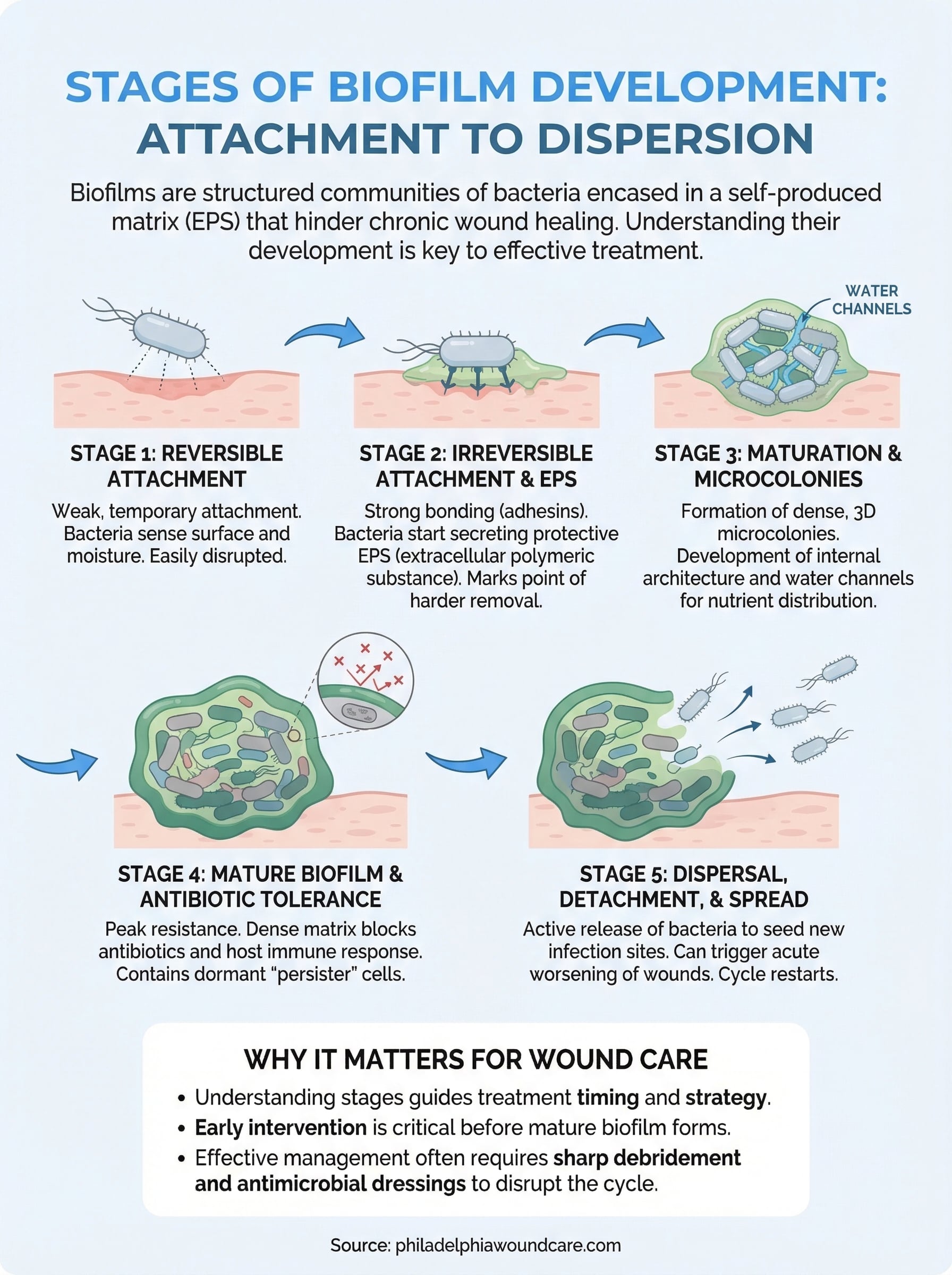
The first stage in the stages of biofilm development begins when a free-floating bacterium makes initial contact with a surface. At this point, the bacterium has not committed to staying. The attachment is weak and temporary, held in place by loose physical and electrostatic forces rather than molecular bonds. A single cell lands on the wound surface and begins sampling its environment, responding to local chemical signals that tell it whether conditions support longer-term colonization. This early contact is the most fragile point in the entire process, and it is where the biofilm is most vulnerable to disruption.
What bacteria are looking for
During reversible attachment, bacteria run a rapid environmental assessment. They respond to surface properties including hydrophobicity, texture, and nutrient concentration. Wound beds offer an appealing target because damaged tissue is rich in host proteins like fibronectin, collagen, and fibrinogen, all of which function as attachment points for bacterial adhesins. Necrotic or sloughy tissue creates particularly favorable conditions because the immune system’s ability to patrol those areas is severely limited by poor blood supply and tissue debris.
Bacteria also use flagella at this stage to actively move toward and across the surface. As they make contact, they begin switching gene expression patterns, starting to downregulate the systems used for free swimming and activating pathways that support surface attachment. This genetic shift is not yet locked in. The bacterium can still reverse course and return to a planktonic state if conditions change, which is exactly what makes this stage so clinically significant.
Mechanical debridement targets the biofilm precisely at this point, physically removing loosely attached bacteria before they produce the EPS matrix that makes later stages far more resistant to treatment.
Why this stage is the critical window for intervention
You cannot see reversible attachment happening with the naked eye, and no bedside test currently identifies it in real time. What clinicians do know is that disrupting bacterial populations early, before they produce EPS and anchor irreversibly, dramatically reduces the overall bacterial burden in a wound. This is why consistent, regular debridement combined with antimicrobial wound dressings remains the foundation of biofilm management even before a mature biofilm has been confirmed.
Waiting until a biofilm is fully established means working against a far stronger biological defense. Acting during or shortly after reversible attachment shifts the odds significantly in your favor.
Stage 2: Irreversible attachment and EPS
When bacteria commit to a surface, the entire dynamic of the wound changes. The transition from reversible to irreversible attachment is triggered by molecular-level bonding between bacterial surface structures and host tissue proteins. Adhesin proteins on the bacterial outer membrane lock onto fibronectin, collagen, and other extracellular matrix components in the wound bed. Once those bonds form, simple mechanical forces are no longer enough to dislodge the bacteria. The window for easy removal has closed.
How EPS production changes the playing field
As bacteria anchor firmly to the surface, they begin secreting the extracellular polymeric substance, or EPS, that defines biofilm structure. This matrix is a complex mixture of polysaccharides, proteins, extracellular DNA, and lipids. The bacteria do not produce it passively. They actively regulate EPS output in response to quorum sensing signals, the chemical communication system that lets bacteria coordinate behavior based on local population density. As more cells arrive and density increases, quorum sensing triggers a coordinated rise in EPS production across the entire colony.
The EPS matrix serves several functions at once. It physically anchors the colony to the wound surface, it creates distinct microenvironments where different bacterial species occupy separate niches, and it begins forming the water channels that will eventually supply nutrients to deeper layers of the mature biofilm. This structural organization is a key reason why the stages of biofilm development progress with such biological efficiency once irreversible attachment occurs.
Once EPS production begins in earnest, standard wound cleaning and irrigation lose most of their effectiveness at clearing bacteria from the wound surface.
Why this stage determines treatment difficulty
The shift to irreversible attachment is the point where clinical intervention becomes substantially harder. Bacteria encased in early EPS already begin demonstrating reduced sensitivity to antimicrobial agents. Topical antiseptics that work well against planktonic bacteria start losing effectiveness because the matrix physically limits their ability to reach bacterial cells. Sharp or mechanical debridement at this stage must be more aggressive than the light cleaning appropriate during reversible attachment, requiring removal of tissue that now harbors an anchored bacterial community.
Your wound care clinician may increase debridement frequency or switch to antimicrobial dressings with biofilm-disrupting agents specifically because of what happens at this second stage. Early, decisive action here directly affects how difficult the mature biofilm becomes to manage in the stages that follow.
Stage 3: Maturation and microcolonies
By the time bacteria reach this third phase in the stages of biofilm development, the colony has moved well beyond a surface layer of anchored cells. Microcolonies, which are dense, three-dimensional clusters of bacteria embedded within growing EPS, begin to organize into recognizable architectural formations. These structures are not random. They form in response to quorum sensing signals and local nutrient gradients, creating a community that distributes workload, shares genetic information, and actively manages resources across the wound surface.
How microcolonies develop internal architecture
As cell density rises within the developing colony, water channels begin forming between and around the microcolony clusters. These channels carry nutrients into the interior of the biofilm and remove metabolic waste products outward, preventing toxic accumulation that would otherwise limit bacterial survival. This internal distribution network is what allows the biofilm to grow significantly thicker and more structurally complex than a simple surface film.
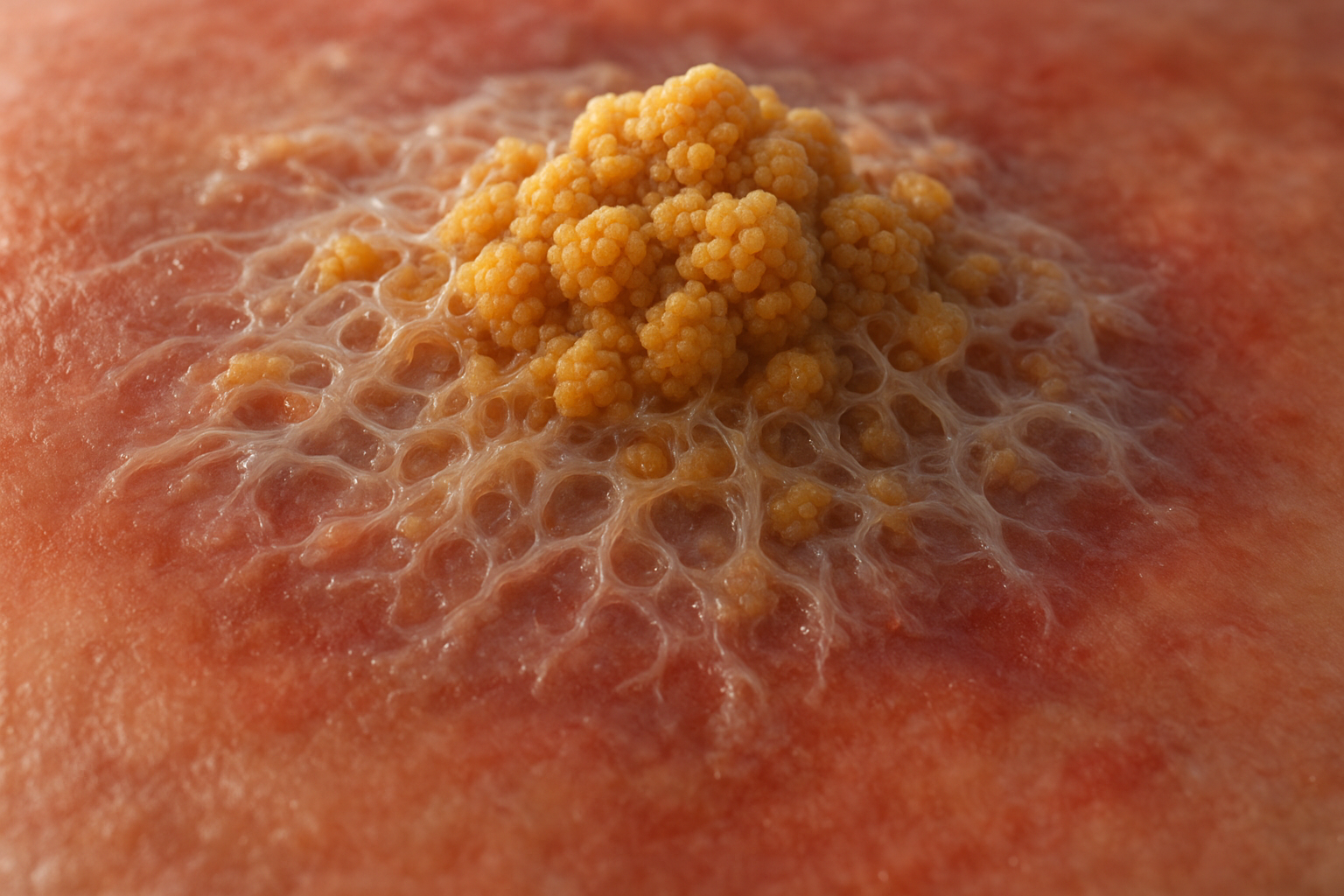
Mature microcolonies within a Stage 3 biofilm can reach several hundred micrometers in thickness, making physical disruption without sharp debridement largely ineffective at this point.
Different bacterial species continue to stratify into distinct spatial niches within the microcolony structure. Aerobic species occupy the outer layers where oxygen is available, while anaerobic species cluster in the deeper, oxygen-depleted zones closer to the wound bed. This stratification means a single biofilm can harbor multiple bacterial populations with entirely different metabolic profiles, each contributing to overall biofilm stability and each requiring a different antimicrobial approach.
What this stage means for your wound’s healing trajectory
At Stage 3, the biofilm is exerting a sustained inflammatory load on the surrounding wound tissue. The colony releases enzymes, toxins, and degradation products that directly damage host cells and break down the growth factors your body needs to rebuild tissue. If your wound has remained stalled for several weeks despite dressing changes and antimicrobial treatment, biofilm maturation at or beyond this stage is a likely contributing factor.
Clinicians managing wounds at this stage typically combine aggressive sharp debridement with biofilm-targeted antimicrobial dressings, often increasing treatment frequency to every one to two weeks. Removing the bulk of the microcolony structure through debridement resets the biofilm to an earlier, more vulnerable stage and gives antimicrobial agents a realistic chance of reaching remaining bacterial cells.
Stage 4: Mature biofilm and antibiotic tolerance
Stage 4 represents the fully developed form of the biofilm, where all the structural and biochemical mechanisms that began in earlier stages of biofilm development have reached their peak functional capacity. The colony now operates as a highly coordinated, self-sustaining community, with established water channels, dense EPS matrix, stratified bacterial populations, and active quorum sensing running continuously across the entire structure. At this point, the biofilm is not simply resistant to treatment; it actively neutralizes many of the standard approaches clinicians use to control wound infection.
How a mature biofilm resists treatment
The EPS matrix in a Stage 4 biofilm is thick, dense, and chemically reactive, capable of physically blocking antimicrobial agents from reaching the bacteria underneath. Beyond simple physical exclusion, the matrix also binds and inactivates certain antibiotics directly, breaking them down before they can penetrate to the bacterial cells. This combination of physical and chemical defense makes the mature biofilm genuinely impenetrable to many agents at standard therapeutic doses.
Research published in the scientific literature consistently shows that bacteria within a mature biofilm can survive antibiotic concentrations 100 to 1,000 times higher than those required to kill the same species in planktonic form.
Bacterial populations within the mature biofilm also include persister cells, a subpopulation that enters a dormant or near-dormant metabolic state. Most antibiotics work by targeting active bacterial processes like cell wall synthesis or DNA replication. Persister cells slow or halt those active processes entirely, making them essentially invisible to antibiotic action. When antibiotic treatment ends, persister cells reactivate and begin repopulating the wound surface.
What antibiotic tolerance means in practice
For your wound care plan, the presence of a mature biofilm explains why prolonged antibiotic courses often produce little measurable improvement in wounds that have stalled for weeks or months. The antibiotic reaches the wound surface but fails to penetrate the EPS matrix in concentrations sufficient to eliminate the bacterial population underneath. Your clinician cannot resolve this by simply increasing the antibiotic dose, because the tolerance mechanisms operating at this stage function independently of concentration.
Effective management at Stage 4 requires combining sharp debridement to physically disrupt and remove the biofilm structure with targeted antimicrobial dressings applied immediately after, before bacteria begin rebuilding. Debridement without immediate antimicrobial follow-up gives bacteria exactly the window they need to re-establish the protective matrix that was just removed.
Stage 5: Dispersal, detachment, and spread
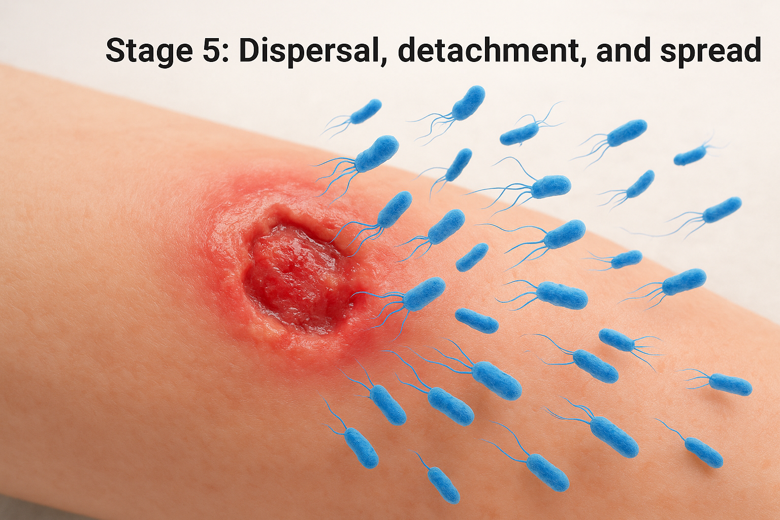
Dispersal is the final stage in the stages of biofilm development, and it is arguably the most dangerous phase for wound management. When a mature biofilm colony reaches capacity or detects unfavorable local conditions, it actively releases bacteria back into a free-floating, planktonic form. These newly released cells do not simply stay in place. They spread to surrounding tissue, seed new infection sites, and begin the entire developmental cycle again at a new location on the wound bed or beyond.
How bacteria trigger dispersal
Biofilm dispersal is not a passive or accidental event. Bacteria within the mature colony actively regulate the decision to detach using quorum sensing signals and environmental cues. When nutrients become depleted, oxygen drops below a functional threshold, or local waste product concentration rises past a critical point, the biofilm community responds by downregulating the genes responsible for EPS production. As the matrix begins to break down from within, flagella expression reactivates in bacteria near the biofilm surface, restoring their ability to swim and allowing them to leave the colony.
Dispersal-phase bacteria express a distinct genetic profile that makes them more virulent and more capable of rapid surface attachment at new sites than standard planktonic bacteria.
Released bacteria enter a planktonic state temporarily, making them briefly more susceptible to antibiotics than their biofilm-protected counterparts. This window is short. Within hours, these cells begin the reversible attachment phase at a new location, restarting the developmental cycle and extending the infection across a wider area of tissue.
What dispersal means for wound management
Dispersal explains a clinical pattern that caregivers and patients often find confusing: a wound that appears to be improving after debridement can suddenly worsen or develop new areas of infection within days of treatment. Debridement disrupts the mature biofilm mechanically, which can inadvertently trigger mass dispersal of viable bacteria across the wound surface before the antimicrobial dressings applied afterward can neutralize them.
Your wound care clinician manages this risk by applying biofilm-targeted antimicrobial dressings immediately after debridement, closing the treatment window as tightly as possible. Leaving even a short gap between debridement and antimicrobial coverage gives dispersing bacteria exactly the conditions they need to initiate a new round of attachment and colony formation, resetting the biofilm cycle and undermining the progress made during treatment.
Key takeaways for patients and caregivers
The stages of biofilm development follow a predictable sequence, and that predictability works in your favor. Early intervention, before bacteria produce their protective EPS matrix, is dramatically more effective than treating a mature, entrenched colony. Every stage from reversible attachment through dispersal represents a distinct clinical challenge, but consistent debridement and immediate antimicrobial coverage after treatment are the two most reliable tools for keeping biofilm from advancing.
Your wound’s stalling or worsening may not reflect anything you are doing wrong. Biofilm-related resistance is a biological process that standard dressings and antibiotics alone cannot overcome. If your wound has stopped progressing despite regular care, you need a clinician who specifically targets biofilm in their treatment approach.
Philadelphia Wound Care brings physician-led wound management directly to your home or care facility. Contact our team to request a mobile wound care evaluation and get a treatment plan built around your wound’s actual needs.