

Granulation Skin Healing: What It Is And What It Means
If you’ve peeked under a bandage and seen bumpy, deep red or pink tissue filling in a wound, you’re looking at granulation skin healing in action. It can look alarming if you don’t know what you’re seeing, almost raw or beefy, and family caregivers often assume it means something has gone wrong. In most cases, it’s the opposite: your body building new tissue exactly where it should.
Granulation tissue is the body’s natural scaffolding, made of new blood vessels and collagen that fill a wound bed before skin can close over it. Its color, texture, and moisture level tell you a lot about whether healing is on track or stalling, which matters most for chronic wounds like diabetic ulcers or pressure sores that don’t follow a typical timeline.
In this article, we’ll walk through what granulation tissue actually is, how it forms during the wound healing stages, and what healthy versus unhealthy granulation looks like. If you’re caring for a loved one with a slow-healing wound, this is the kind of information that helps you know when to relax and when it’s time to call for a specialist’s evaluation.
Why granulation tissue matters for wound healing
Granulation as the bridge between injury and closure
Granulation tissue forms during the proliferative phase of wound healing, the stage right after your body has cleared out dead tissue and debris. Without it, a wound simply can’t close. Skin cells (keratinocytes) need something to migrate across, and granulation tissue provides that surface. Think of it as scaffolding, not the final structure. It fills the wound bed with new capillaries and collagen so that skin cells have solid ground to travel over as they knit the edges back together. When this stage stalls, everything downstream stalls with it, which is why clinicians watch granulation so closely in chronic wounds.
The four stages of wound healing at a glance
Understanding where granulation fits helps explain why its appearance is such a useful checkpoint. Most wounds move through four overlapping phases, though chronic wounds often get stuck in one for weeks or months instead of days.
| Stage | What’s happening | Typical timeframe |
|---|---|---|
| Hemostasis | Blood clots form to stop bleeding | Minutes to hours |
| Inflammation | Immune cells clear debris and bacteria | 1 to 5 days |
| Proliferation (granulation) | New blood vessels and collagen fill the wound bed | 5 days to several weeks |
| Maturation | Skin cells cover the wound and tissue remodels | Weeks to months |
For a healthy adult with a minor cut, this sequence runs smoothly on its own. For someone with diabetes, poor circulation, or a pressure injury from limited mobility, the proliferation stage can drag on far longer than it should, and that’s where problems tend to surface.
What happens when granulation stalls
Stalled or absent granulation isn’t just a cosmetic delay. It’s a signal that the underlying biology of healing has broken down somewhere. Wounds that sit without adequate granulation tissue stay open longer, which raises the risk of infection and gives bacteria time to form biofilm, a protective layer that makes germs harder to treat with standard antibiotics or topical antiseptics. According to the Centers for Disease Control and Prevention, chronic wounds affect millions of Americans each year, many of them older adults with diabetes or vascular disease, and delayed healing is a major driver of hospital readmissions in this group.
Granulation tissue is your body’s proof that a wound is actively rebuilding itself, and its absence is often the clearest early sign that something needs medical attention.
Recognizing that connection early matters more than most caregivers realize. A wound that looks "stuck" at the same size and depth for two or three weeks, with little visible granulation, usually needs a clinical evaluation rather than more time and patience. This is especially true for pressure ulcers and diabetic foot wounds, where poor blood flow or nerve damage can mask pain signals that would otherwise prompt someone to seek help sooner. The Agency for Healthcare Research and Quality notes that pressure injuries alone contribute to significant morbidity in long-term care settings, underscoring why tracking granulation isn’t just academic. It’s a practical, visual way to catch a stalling wound before it becomes a bigger medical problem.
How to tell if your granulation tissue is healthy
The look and feel of healthy tissue
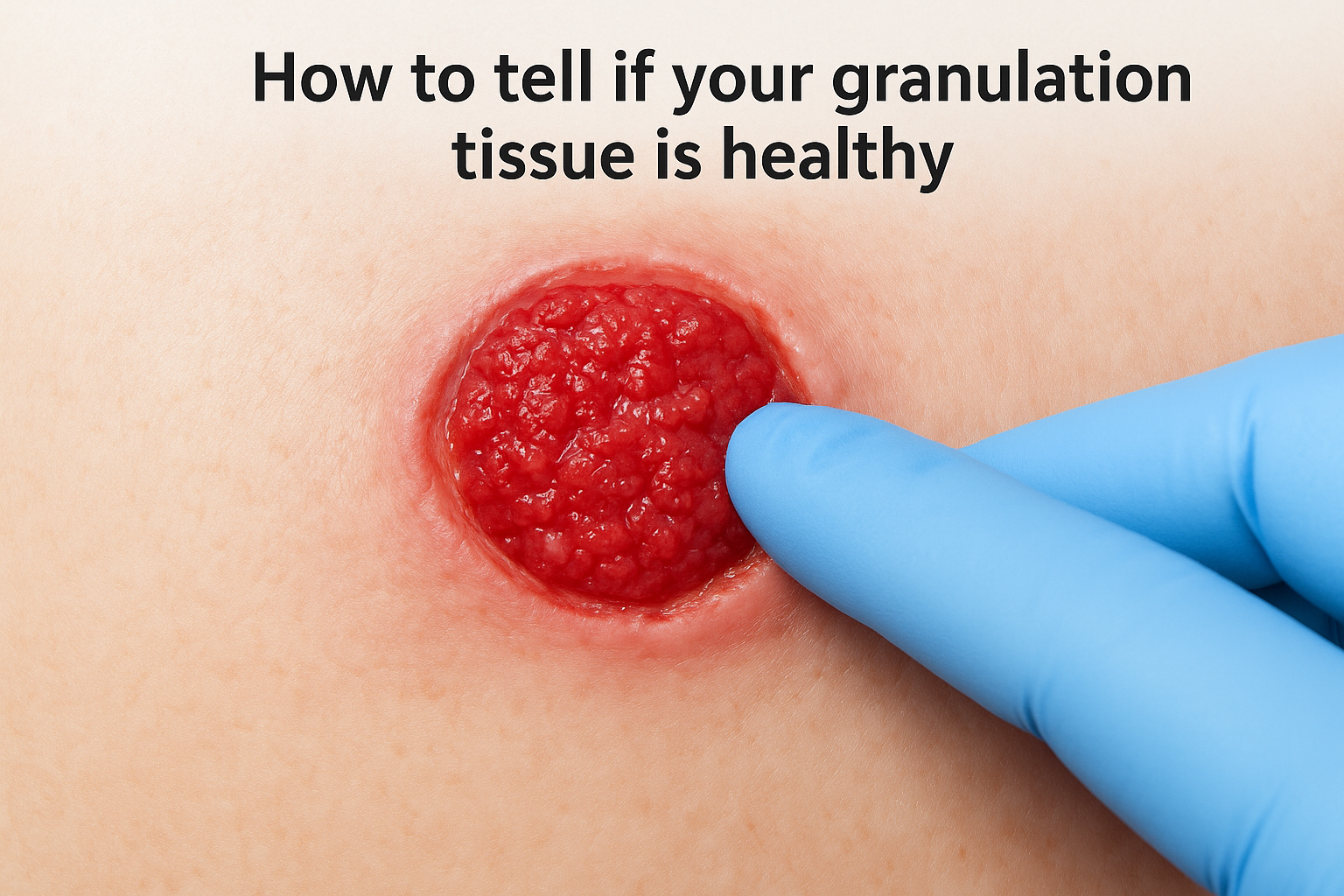
Healthy granulation tissue has a specific appearance that’s easy to recognize once you know what to look for. It should be beefy red or deep pink, moist without being soupy, and slightly bumpy, almost like the surface of a raspberry. Touch isn’t usually part of a caregiver’s job, but clinicians often note that healthy granulation bleeds a little with minor contact, which is actually a good sign of strong blood supply. Texture matters as much as color here: a wound bed that looks glassy, dry, or unusually smooth often isn’t building the vascular network it needs.
Reading color as a health indicator
Color tells you more than most caregivers expect, and it’s the fastest visual check you can do at each dressing change. Pale pink or dusky tissue can point to poor circulation or early infection, while dark red or maroon patches sometimes signal trauma from dressings that are too tight or changed too roughly. Yellow, gray, or white patches usually mean the tissue isn’t granulation at all, but slough or dead tissue that needs to be addressed before healing can move forward.
A healthy wound bed should look alive, glistening, red, and slightly uneven, never flat, pale, or slick.
A quick reference for what you’re seeing
Use this table as a fast gut check during dressing changes. It won’t replace a clinical exam, but it gives you language to describe what you’re seeing when you call a physician or nurse.
| What you see | Likely meaning |
|---|---|
| Deep red, moist, slightly bumpy | Healthy granulation, healing on track |
| Pale pink, dry surface | Poor blood flow, slowed healing |
| Dark maroon or purple | Bruising, pressure damage, or fragile new vessels |
| Yellow or tan film | Slough, dead tissue blocking granulation |
| Grayish or black tissue | Necrotic tissue, needs prompt medical evaluation |
| Shiny, overly smooth surface | Excess moisture or hypergranulation |
Size and shape as healing clues
Granulation should also fill in gradually from the edges and base of the wound, shrinking the overall size week over week. If the wound bed looks the same size and depth at two consecutive dressing changes, that’s worth flagging even if the color looks acceptable. Odor, drainage color, and surrounding skin temperature round out the full picture, but color and texture remain the two fastest signals for anyone without formal training to track between visits from a home health nurse or wound care physician.
How to support healthy granulation at home
Keep the wound bed moist but not wet
Moisture balance drives healthy granulation more than any single product on a shelf. Moist wound healing keeps new blood vessels and collagen from drying out, while too much drainage softens the surrounding skin and invites breakdown. Stick to the dressing type and change schedule your wound care team sets, since swapping products on your own can throw off a balance that’s already working in your favor.
Consistent, moist, protected wound care does more for granulation than any single ointment or supplement ever will.
Feed the healing process with the right nutrition
Nutrition supplies the raw materials granulation tissue actually needs to build itself. Protein intake matters most, since collagen production depends on it, and older adults recovering from chronic wounds often need more protein than they’re used to eating. Steady blood sugar control matters just as much for anyone managing diabetes, because elevated glucose slows collagen synthesis and blunts circulation right at the wound bed.
- Aim for lean protein at most meals: eggs, poultry, fish, beans, or a protein supplement if appetite is low
- Keep blood sugar within the range your physician has recommended
- Drink water consistently through the day rather than in one large amount
- Ask your care team about vitamin C or zinc if a deficiency is suspected
Protect the site between dressing changes
Pressure and friction remain the biggest at-home threats to granulation tissue that’s already forming. Repositioning a bedbound loved one every two hours, using pressure-relief cushions, and keeping heels off the mattress all reduce the mechanical stress that can reopen fragile new tissue. Offloading footwear for diabetic foot ulcers works the same way, shifting weight off the healing area so new capillaries aren’t crushed with every step.
Watch for what not to do
Skipping dressing changes isn’t the only mistake caregivers make around a healing wound. Scrubbing the area with soap, applying over-the-counter antiseptics the care team hasn’t approved, or leaving a dressing on days past its recommended change can all interfere with delicate granulation tissue. When in doubt, call the physician’s office before trying a new product, even one marketed for wound care, since some ingredients are simply too harsh for tissue this fragile.
Warning signs that need professional attention
Color changes that point to infection
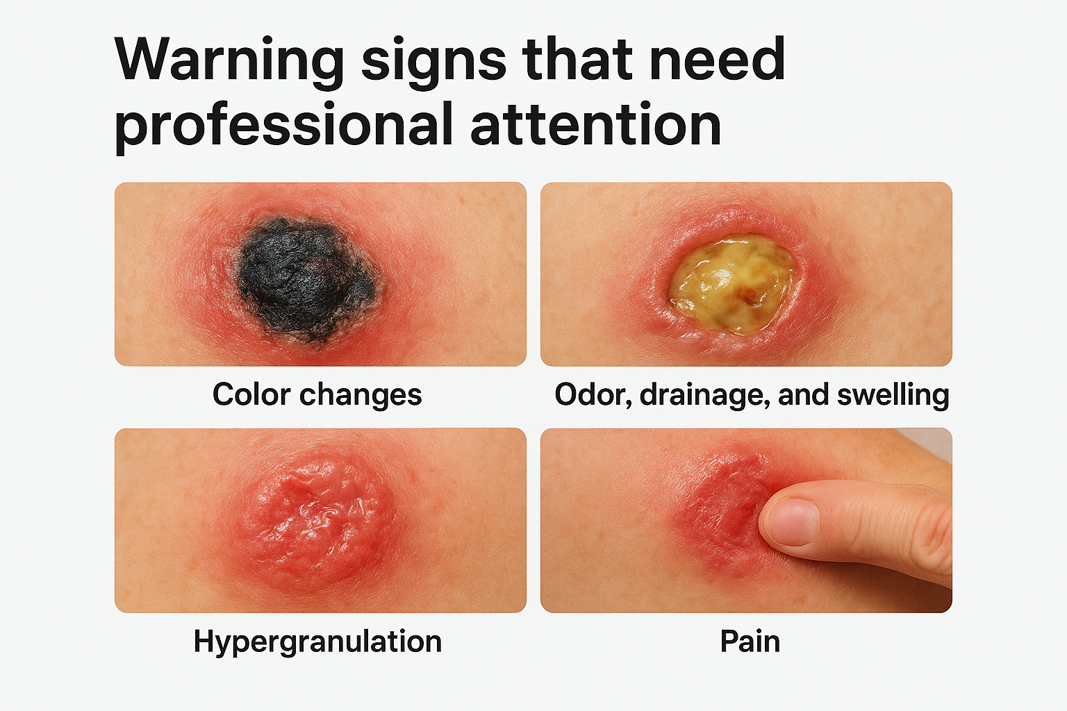
Spotting a shift from healthy red tissue to something darker or duller is often the first clue that a wound has taken a turn. Necrotic tissue that looks black, gray, or dark brown means cells have died and the body can’t build granulation over dead material, no matter how good your dressing technique is. Greenish or unusually yellow patches, especially paired with a foul smell, often point toward bacterial infection rather than normal healing debris. None of these changes are something to monitor at home for another week; they call for a same-day evaluation.
Odor, drainage, and swelling that don’t fit the pattern
Drainage tells its own story, and a sudden change in volume, color, or thickness usually means something is off underneath the surface. Thick yellow or green fluid, a strong or sour smell, or drainage that soaks through a dressing faster than it used to are all signs of possible infection. Increased swelling around the wound edges, warmth that spreads beyond the immediate area, or new redness creeping outward all suggest the body is fighting something more than routine tissue repair. Fever in an elderly patient, even a low-grade one, should never be dismissed as unrelated to a wound that looked stable a few days earlier.
Any wound that suddenly smells worse, drains more, or triggers a fever needs same-day medical attention, not a wait-and-see approach.
Hypergranulation: too much of a good thing
Granulation tissue can also become a problem when it grows past the wound edges instead of stopping level with the surrounding skin. This is called hypergranulation, and it shows up as tissue that looks shiny, raised, and sometimes almost bubbly. It’s more common under occlusive dressings left on too long or in wounds with ongoing moisture imbalance. Left alone, hypergranulation can block skin cells from migrating across the wound bed and closing it properly, so it usually needs a clinician’s assessment rather than a dressing change alone.
Pain that doesn’t match what you see
Questioning a sudden pain change matters just as much as watching color and drainage. A wound that becomes noticeably more painful, especially in patients with diabetic neuropathy who normally feel little at all, can signal deep tissue involvement or infection tracking below the surface. Use this quick checklist to decide when a phone call can’t wait:
- Fever above 100.4°F alongside wound changes
- New or spreading redness beyond the wound border
- Foul odor or a sudden increase in drainage
- Black, gray, or unusually dark tissue in the wound bed
- Rapidly worsening pain, especially in someone with reduced sensation
When to call in a mobile wound care physician
Deciding between a wait-and-see approach and a house call gets easier once you know what a mobile physician actually brings to the bedside. Physician-led evaluation at home means a licensed surgeon examines the wound in person, often within 24 hours of a referral, rather than a caregiver trying to describe color and drainage over the phone to an office that can’t see what’s happening.
Why a house call beats another trip to urgent care
Getting an elderly or mobility-impaired patient to a clinic appointment is often harder than the wound itself. Transportation, wheelchair access, and the physical strain of travel can delay care by days, and every day a stalled wound goes unassessed raises the risk of infection or deeper tissue breakdown. A mobile wound care physician removes that barrier entirely by bringing the exam, the treatment plan, and follow-up care directly to a private residence, skilled nursing facility, assisted living community, or hospice setting.
Getting expert eyes on a wound within a day matters more than which building the visit happens in.
What a home visit typically includes
A house call for a stalled or worsening wound isn’t a quick glance and a new bandage. It usually covers a full clinical assessment along with next steps for advanced treatment options that go beyond standard dressings.
- In-person wound measurement, debridement if needed, and infection screening
- Discussion of allograft or regenerative therapy for wounds not responding to standard care
- Coordination with the referring physician, facility nurse, or case manager on an updated care plan
- Direct billing coordination with Medicare, Medicare Advantage, or commercial insurance
Signs it’s time to make the call
Any of the warning signs covered earlier, spreading redness, foul odor, new pain, or a wound bed that hasn’t changed in two weeks, are reason enough to request an evaluation rather than waiting for the next scheduled visit. Facility staff and family caregivers alike benefit from a rapid response system that doesn’t require an ER trip just to get a specialist’s opinion on a slow-healing ulcer.
Referring physicians and case managers also lean on mobile wound care as a bridge after hospital discharge, when a patient’s wound needs closer follow-up than a standard home health nurse visit can provide. That continuity, from hospital bed to full closure, is exactly where a physician-led mobile practice earns its place in the recovery timeline.
Keeping healing on track
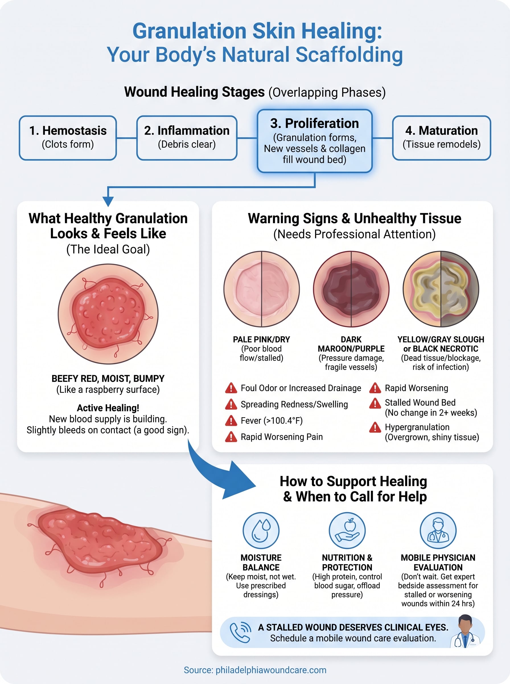
Granulation tissue is your best visual clue that a wound is healing the way it should. Beefy red, moist, slightly bumpy tissue means new blood vessels and collagen are doing their job. Pale, dry, dark, or overgrown tissue means something needs attention before the wound can close.
Knowing the difference gives you real confidence at every dressing change, whether you’re a family caregiver or facility staff managing a slow-healing ulcer. But visual checks at home only go so far. A stalled wound bed, spreading redness, or a change that just doesn’t sit right deserves a clinical set of eyes, not another week of watching and waiting.
If a loved one’s wound has stopped progressing or shows any of the warning signs covered above, don’t wait for it to worsen. Schedule a mobile wound care evaluation and get a physician’s assessment at the bedside, often within 24 hours.