
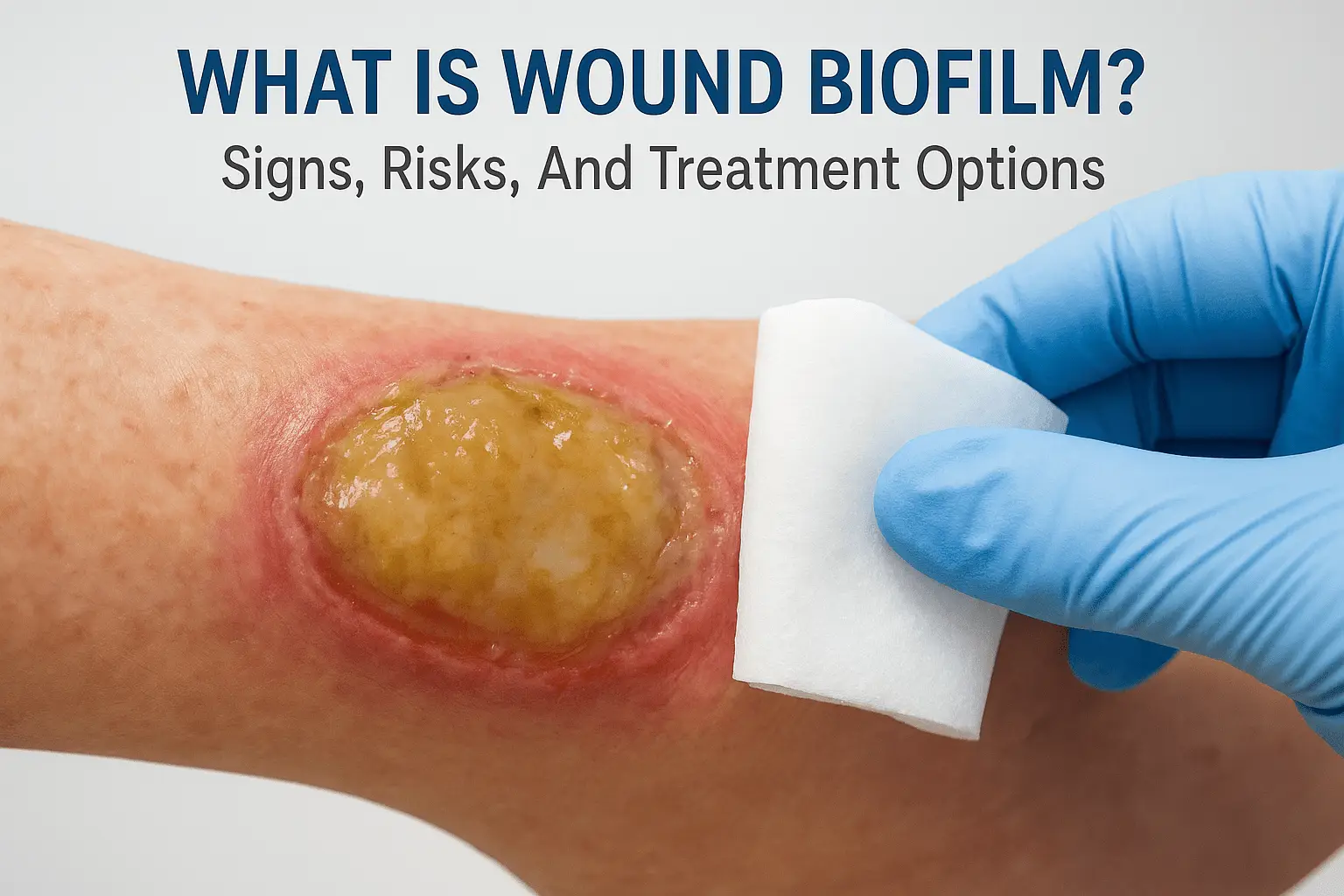
What Is Wound Biofilm? Signs, Risks, And Treatment Options
If you’ve been caring for a chronic wound that refuses to improve despite consistent treatment, a hidden layer of bacteria could be the reason. Understanding what is wound biofilm matters because this microscopic barrier is one of the most common, and most overlooked, obstacles to healing. Biofilm forms when bacteria band together on a wound’s surface, shielding themselves behind a protective matrix that resists antibiotics and the body’s own immune defenses. The result is a wound stuck in a cycle of persistent inflammation with little to no progress toward closure.
Biofilm affects an estimated 60–80% of chronic wounds, including diabetic ulcers, pressure injuries, and venous leg ulcers. For patients who are elderly, homebound, or managing multiple health conditions, the consequences of undetected biofilm go beyond delayed healing, they raise the risk of serious infection and hospitalization.
At Philadelphia Wound Care, our physician-led mobile practice brings advanced wound assessment and treatment directly to patients at home, in skilled nursing facilities, and in hospice settings across the Philadelphia area. Identifying and managing biofilm is a core part of the specialist care we provide at the bedside. This article explains what biofilm is, how to recognize its signs, why it stalls wound healing, and what treatment options are available to get recovery back on track.
What wound biofilm is and how it forms
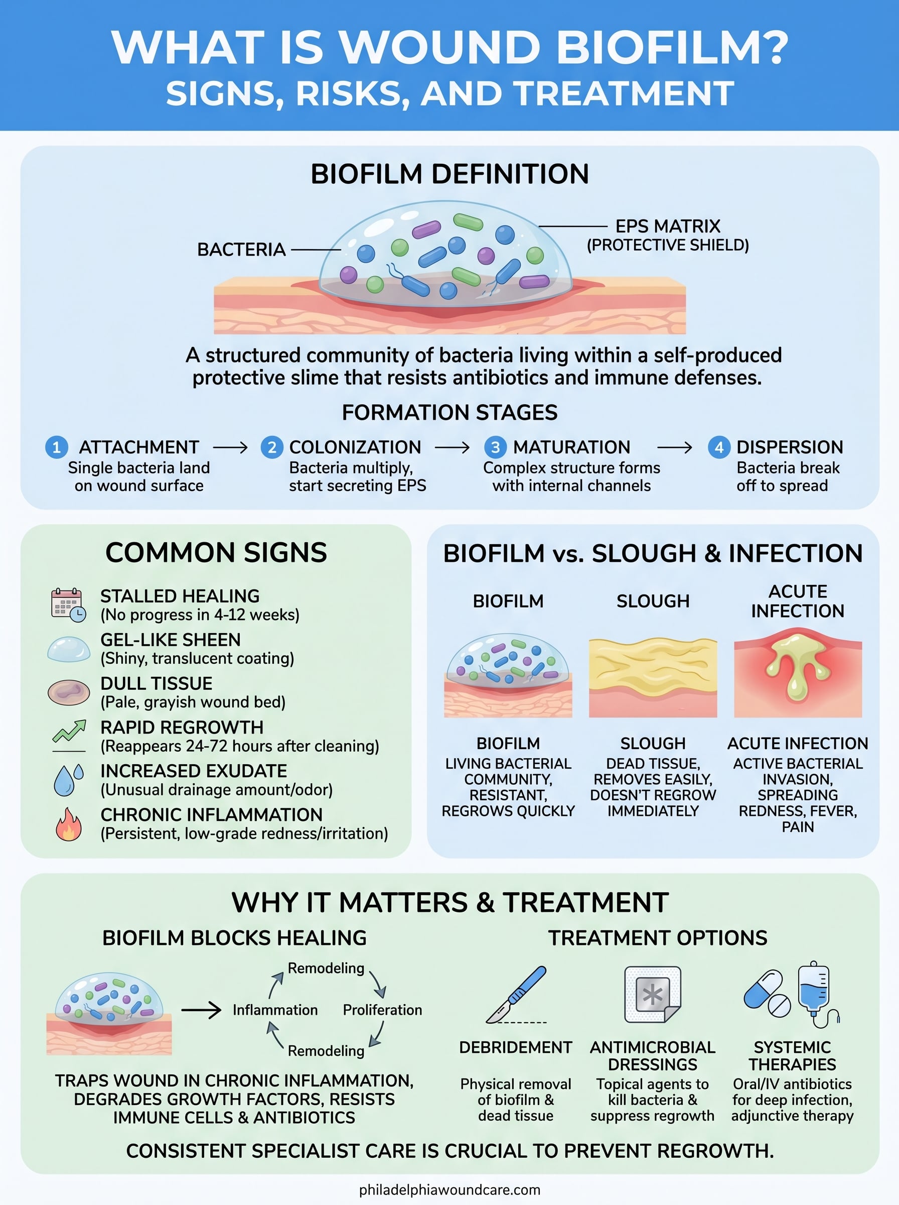
Understanding what is wound biofilm starts with recognizing that bacteria rarely act alone. When bacteria land on a wound surface, they do not simply sit there as isolated cells. Instead, they attach, multiply, and organize into structured communities that behave very differently from free-floating bacteria. These communities anchor themselves to the wound bed and then produce a sticky, gel-like substance called an extracellular polymeric substance (EPS) matrix. That matrix is the defining feature of a biofilm, wrapping the bacterial colony in a physical shield that blocks many of the treatments designed to kill it.
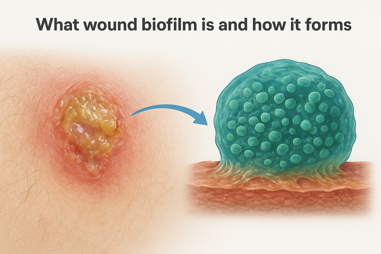
The bacterial community behind biofilm
A wound biofilm is not a single species working alone. Multiple types of bacteria, including Staphylococcus aureus, Pseudomonas aeruginosa, and Enterococcus, frequently coexist within the same biofilm. This mixed-species environment creates what researchers call a polymicrobial community, where different bacteria support each other’s survival. Some species break down antibiotics before they reach neighboring bacteria, while others produce compounds that suppress your immune response. The result is a collective defense system far stronger than any individual bacterial strain could build on its own.
A single wound biofilm can contain dozens of bacterial species working in coordination, making it significantly harder to treat than a conventional single-strain infection.
The stages of biofilm development
Biofilm does not appear overnight, but it forms faster than many people expect. The process moves through distinct developmental stages, each one making the biofilm progressively harder to remove.
The development follows a recognizable sequence:
- Attachment: Free-floating bacteria make initial contact with the wound surface and begin adhering to the tissue within hours of exposure.
- Colonization: Attached bacteria multiply and start secreting the EPS matrix, anchoring the community more firmly to the wound bed.
- Maturation: The biofilm grows in depth and complexity, developing internal channels that distribute nutrients and oxygen through the colony.
- Dispersion: Portions of the mature biofilm break off, releasing bacteria that can spread to new areas of the wound or nearby tissue and restart the entire cycle.
This cycle can restart within 24 to 72 hours after a biofilm is disrupted, which is one key reason a single debridement session is rarely enough to resolve a chronic biofilm problem on its own.
Why the protective matrix is so difficult to penetrate
The EPS matrix is not just a passive shell. It actively slows the diffusion of antibiotics into the biofilm core, giving interior bacteria time to adapt or survive exposure. The matrix also neutralizes enzymes produced by your immune system before they can reach the bacteria underneath. Research accessible through the National Institutes of Health has shown that bacteria living inside a mature biofilm can be up to 1,000 times more resistant to antibiotics than the same bacteria in a free-floating state.
Your body’s white blood cells, which normally engulf and destroy bacteria, struggle to penetrate the biofilm matrix as well. They detect the biofilm’s presence and respond by releasing inflammatory signals, but those signals cannot clear the infection. Instead, they sustain a chronic inflammatory state in the wound that damages surrounding tissue and blocks the normal progression toward healing. This persistent, low-grade inflammation is one of the clearest indicators that a biofilm may be present, even when no obvious signs of a classical infection, such as fever or spreading redness, are visible to a caregiver or even a general clinician.
Why wound biofilm matters in chronic wounds
Chronic wounds are wounds that fail to progress through the normal stages of healing within four to twelve weeks. They include diabetic foot ulcers, venous leg ulcers, pressure injuries, and arterial ulcers. Understanding what is wound biofilm becomes especially important in this context because chronic wounds provide exactly the kind of environment where biofilm thrives: low oxygen levels, compromised blood supply, and an already-weakened immune response. Biofilm does not cause every chronic wound, but its presence is a major reason so many chronic wounds resist even well-executed standard care.
Why chronic wounds are particularly vulnerable
Patients managing diabetes, peripheral vascular disease, or heart failure often have reduced circulation to their extremities. Poor blood flow limits the delivery of immune cells and oxygen to the wound site, which slows the body’s ability to detect and fight bacterial colonization before it progresses. By the time biofilm has matured in a wound with compromised circulation, the protective EPS matrix is already well-established, making it far harder to treat with topical antimicrobials or oral antibiotics alone.
Older adults face additional risk because immune function naturally declines with age, reducing the speed and strength of the initial response to bacterial colonization. When you combine impaired immunity with limited mobility and skin that is thinner and more fragile, the conditions for persistent biofilm are almost ideal. This is why biofilm is disproportionately common in the elderly and homebound populations that Philadelphia Wound Care serves.
The broader health consequences of untreated biofilm
When biofilm goes undetected or undertreated in a chronic wound, the ongoing inflammatory response it triggers does not stay contained to the wound bed. Prolonged wound inflammation raises the overall bacterial burden in the tissue, increasing the risk of spreading cellulitis, osteomyelitis (bone infection), and systemic sepsis. For patients who are already medically fragile, any one of these complications can lead to hospitalization, surgical intervention, or worse outcomes.
Studies in wound care literature consistently show that wounds with confirmed biofilm take significantly longer to heal and carry a higher risk of amputation in diabetic patients compared to wounds without biofilm.
Beyond the physical risks, untreated biofilm places a substantial burden on caregivers and healthcare systems. Wound dressings need to be changed more frequently, specialist visits become more frequent, and the psychological toll on patients who see no progress compounds over time. Recognizing biofilm early and treating it with the right clinical approach is the most direct way to break that cycle.
Common signs and symptoms of wound biofilm
Biofilm rarely announces itself with the classic warning signs of infection you might expect. There is typically no fever, no spreading redness, and no obvious pus that would prompt an immediate call to a physician. Instead, recognizing what is wound biofilm in a real wound means paying attention to subtler, persistent patterns that standard wound care fails to resolve. Knowing these signs helps you communicate more clearly with your clinical team and push for the right level of assessment before the wound deteriorates further.
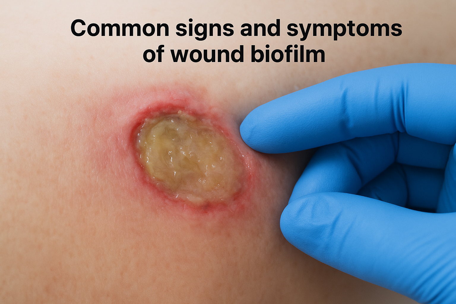
The absence of classic infection symptoms does not rule out biofilm. Many wounds carrying a significant biofilm burden look deceptively stable while making little to no healing progress.
Visual signs on the wound surface
The wound bed itself often provides the clearest early clues. A dull, gel-like sheen on the wound surface is one of the most frequently reported visual indicators of biofilm presence. The tissue may look glassy or slightly translucent rather than the healthy red-pink color of granulating tissue. Some wounds display a surface film that is difficult to wipe away cleanly, returning quickly after removal, which reflects the biofilm’s ability to re-establish itself rapidly after surface disruption.
Common visual indicators to look for include:
- A persistent slippery or gel-like coating on the wound bed
- Tissue that appears pale, grayish, or fails to develop healthy granulation
- A wound surface that looks the same week after week despite regular dressing changes
- Minimal or absent signs of new tissue growth at the wound margins
Physical characteristics and wound behavior
Beyond what you can see, biofilm-associated wounds behave differently from wounds that are simply slow to heal due to circulation or nutrition problems alone. One of the most telling patterns is a wound that responds briefly to a treatment, such as antimicrobial dressings or topical antibiotics, then regresses to its previous state within days. This cycle of temporary improvement followed by stagnation is a strong behavioral marker of biofilm activity.
The wound may also produce more exudate than expected for its size, with drainage that has an unusual consistency or odor that is difficult to attribute to a specific identifiable cause. Surrounding skin can show mild irritation or maceration without a clear reason. If you notice that your wound care routine produces short-lived results and the wound repeatedly returns to the same unresolved state, raising the possibility of biofilm with a wound care specialist is a reasonable and important next step.
Wound biofilm vs slough and regular infection
When clinicians assess a chronic wound, they frequently encounter three different problems that can look similar at first glance: slough, a conventional bacterial infection, and biofilm. Distinguishing between them matters because each one requires a different clinical response. Treating a biofilm as if it were ordinary slough or a standard infection leads to the same outcome you have likely already seen: temporary improvement followed by a wound that stalls all over again. Understanding what is wound biofilm in contrast to these other wound findings helps you ask the right questions when speaking with your care team.
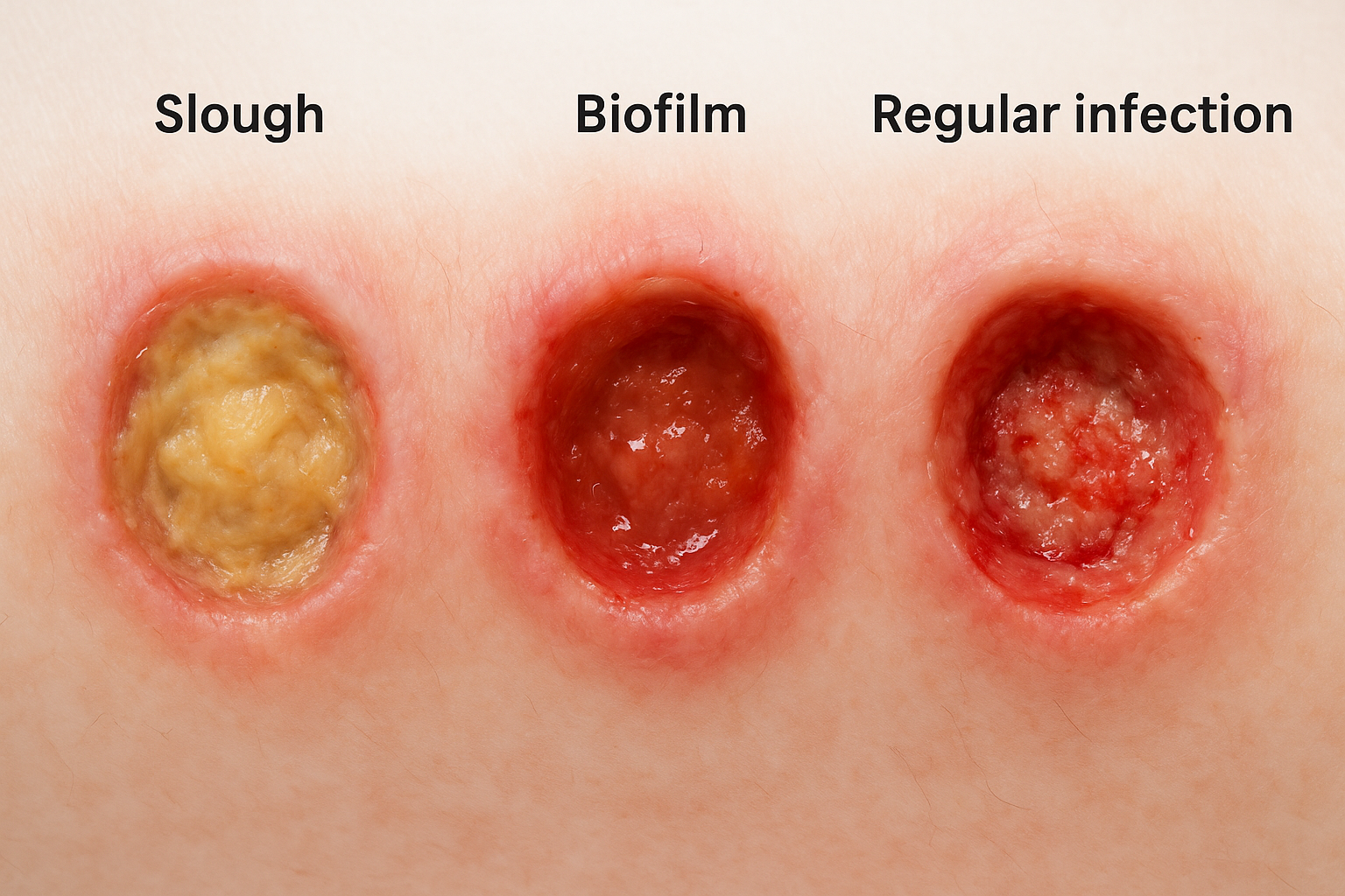
How slough differs from biofilm
Slough is devitalized, non-living tissue that accumulates on a wound surface during normal wound breakdown. It typically appears as a soft, stringy, yellow or tan material that lifts away with debridement and does not return immediately. Slough is part of the body’s natural process of shedding dead cells, and removing it consistently usually allows the wound bed to progress toward healing. Biofilm, by contrast, is a living bacterial structure that returns to the wound surface within 24 to 72 hours after removal. If you notice a gelatinous coating on the wound that reappears quickly after cleaning or dressing changes, that behavior points toward biofilm rather than straightforward slough.
Slough that keeps coming back at the same pace despite consistent debridement should prompt a clinical reassessment for biofilm involvement.
How regular infection differs from biofilm
A conventional acute wound infection involves bacteria multiplying in tissue and triggering a strong inflammatory response that your body and clinicians can usually detect through clear, recognizable signs. These signs include spreading redness around the wound margins, increased warmth, swelling, purulent drainage, and often systemic symptoms like fever or elevated white blood cell count. Standard wound cultures frequently identify the responsible pathogen, and a targeted course of antibiotics generally resolves the problem over days to a few weeks.
Biofilm infections behave very differently. They produce a low-grade, chronic inflammatory state rather than the acute response associated with conventional infection. Your body detects the bacterial presence and sends immune cells to the site, but the EPS matrix prevents those cells from clearing the bacteria. Standard wound cultures often miss biofilm-forming bacteria because they detect free-floating cells, not bacteria embedded in the protective matrix. This means a wound culture returning with negative or insignificant results does not rule out biofilm as a factor. Recognizing this distinction is one of the most important reasons to pursue physician-led specialist evaluation when a chronic wound fails to improve on a conventional treatment plan.
Why biofilm makes wounds hard to heal
Understanding what is wound biofilm clarifies why it causes such persistent treatment failures. A wound moves through an orderly sequence of healing phases: hemostasis, inflammation, proliferation, and remodeling. Each phase depends on the previous one completing successfully. Biofilm actively disrupts this sequence by holding the wound in the inflammatory phase indefinitely, preventing it from advancing to the tissue-building stages where real progress happens. The longer a biofilm persists, the more entrenched this arrested healing state becomes, and the harder it is to restart forward momentum without specialist intervention.
Biofilm sustains a destructive inflammatory loop
When your immune system detects biofilm on a wound surface, it releases inflammatory signals called cytokines and proteases in an attempt to clear the bacteria. The problem is that the EPS matrix blocks immune cells from reaching the bacteria underneath, so the inflammatory response keeps firing without resolving anything. These proteases, which your body produces to fight infection, end up breaking down collagen and growth factors that healthy tissue needs to repair itself. The wound bed is caught between an immune system that cannot finish the job and a bacterial community that cannot be cleared through inflammation alone.
Prolonged protease activity in a biofilm-affected wound actively destroys the building blocks your body needs to close the wound, turning the healing response into part of the problem.
Biofilm degrades the wound environment over time
Your body relies on growth factors like vascular endothelial growth factor (VEGF) and fibroblast growth factor (FGF) to stimulate new blood vessel formation and tissue production. Biofilm and the inflammatory environment it creates reduce the availability of these signals, leaving the wound with insufficient biological instruction to build new tissue. Research has shown that wounds with active biofilm contain measurably lower concentrations of functional growth factors compared to wounds progressing normally toward closure.
New granulation tissue, the healthy, vascularized tissue that fills a healing wound from the base, forms poorly or not at all in a wound environment dominated by biofilm. Any new tissue that does begin to develop is vulnerable to damage from ongoing protease activity. This combination of impaired growth factor signaling and active tissue breakdown explains why so many chronic wounds appear to plateau, showing no measurable improvement in wound dimensions or depth despite weeks of regular dressing changes and topical treatments.
How clinicians diagnose suspected biofilm
Diagnosing biofilm is not straightforward because no single bedside test confirms its presence with certainty. Clinicians instead rely on a combination of clinical observation, wound behavior patterns, and, where available, specialized laboratory analysis. When you work with a wound care specialist rather than a general practitioner, they approach this diagnostic process systematically, using the wound’s history and physical characteristics as the primary guide before ordering any additional testing.
Clinical assessment and visual examination
A wound care physician begins by reviewing how the wound has responded to previous treatments and how long it has been stalled. A wound that plateaued despite appropriate standard care for four or more weeks, with no measurable improvement in size or depth, raises immediate suspicion for biofilm involvement. During the physical examination, the clinician looks for the gel-like surface coating, dull wound bed appearance, and absence of healthy granulation tissue described in the signs section above.
The pattern of wound behavior carries significant diagnostic weight. If debridement or antimicrobial dressings produce brief improvement followed by rapid regression, that cycle is one of the strongest clinical indicators a specialist will use to guide the diagnosis. Your wound’s full treatment history helps the clinician distinguish biofilm-related stagnation from healing delays caused by underlying factors like poor circulation or nutritional deficiency.
Clinical diagnosis of biofilm relies heavily on recognizing patterns over time, not just what a wound looks like at a single visit.
Why standard wound cultures often miss biofilm
Routine wound swab cultures are designed to detect free-floating, planktonic bacteria circulating on the wound surface. Because biofilm bacteria are embedded within the EPS matrix and behave differently from free-floating cells, a standard culture frequently returns negative or insignificant results even when a mature biofilm is present. This is one reason a wound can receive repeated courses of antibiotics with little lasting effect while the culture report gives no clear explanation for treatment failure.
Advanced diagnostic approaches
When clinical suspicion is high, specialized techniques provide stronger evidence. Confocal laser scanning microscopy and fluorescence imaging can visualize bacterial communities directly within wound tissue samples, confirming biofilm structure and distribution. Some facilities use quantitative tissue biopsy, where a sample taken from the wound bed is processed to measure bacterial load at a level that surface swabs cannot capture.
Fluorescence-based wound imaging devices, increasingly available to mobile and specialist practices, allow clinicians to detect bacterial fluorescence patterns at the point of care, giving a real-time picture of bacterial burden without waiting for laboratory results. These tools make earlier, more accurate identification of biofilm possible, particularly for patients receiving care in settings away from a traditional wound clinic.
Treatment options to remove and control biofilm
Once you understand what is wound biofilm and how it behaves, effective treatment follows a logical strategy: disrupt the biofilm physically, reduce the bacterial burden, and sustain that reduction long enough for healing to restart. No single product or intervention accomplishes all three goals alone. Successful biofilm management requires a coordinated approach that your wound care physician selects based on the wound’s location, severity, and the patient’s overall health status.
Debridement as the first line of attack
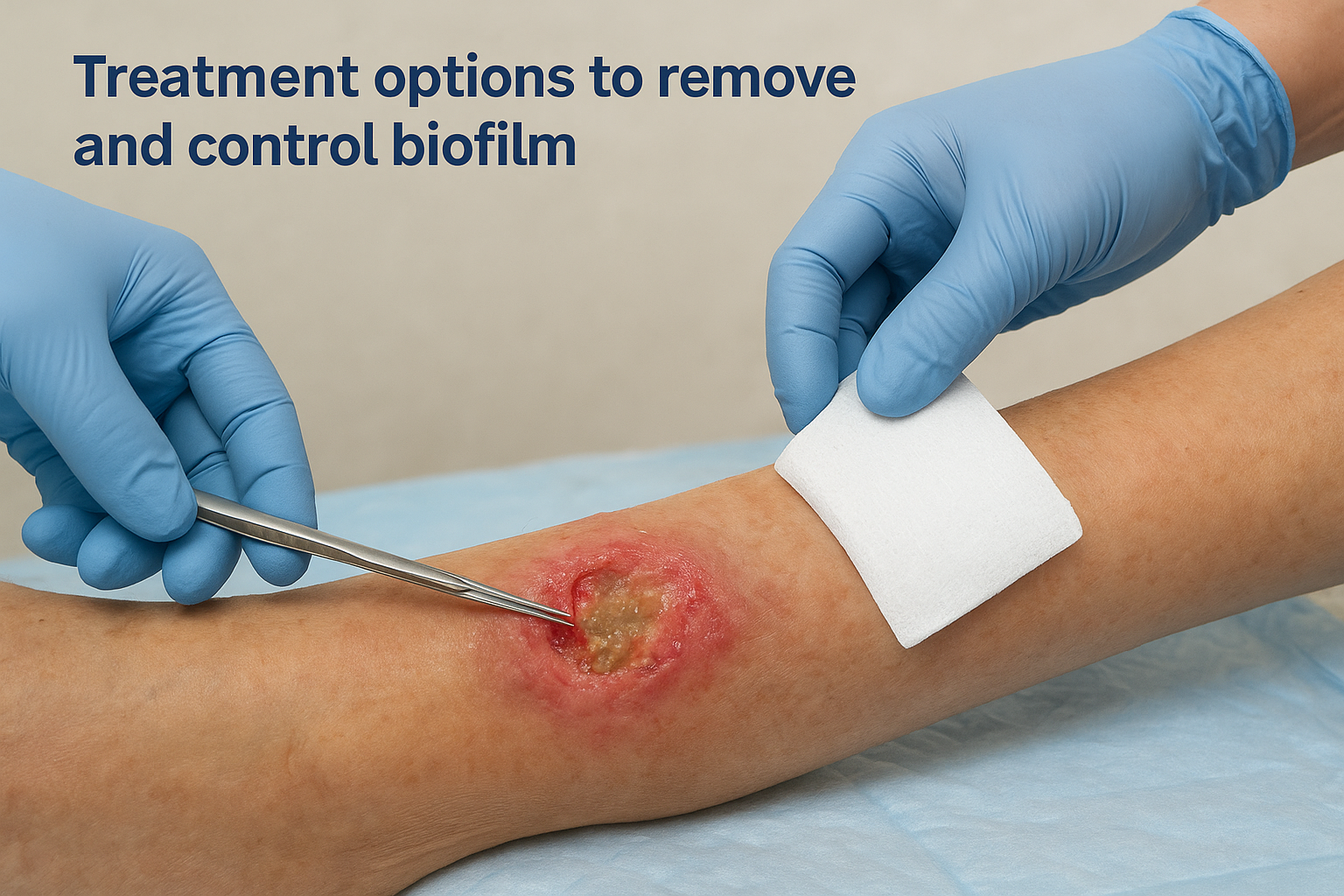
Debridement, the physical removal of non-viable tissue and biofilm material from the wound bed, is the most direct and essential step in breaking down an established biofilm. Sharp debridement, performed by a trained clinician using a scalpel or curette, removes the bulk of the biofilm structure immediately and disrupts the EPS matrix that protects the bacteria underneath. This mechanical disruption reduces the bacterial burden significantly and temporarily resets the wound environment, creating a window for other treatments to work more effectively.
Debridement alone does not eliminate biofilm permanently. Repeating it consistently, often at each clinical visit, is what prevents the biofilm from re-establishing at its previous density.
Autolytic and enzymatic debridement options, delivered through specialized dressings and topical agents, support ongoing tissue breakdown between clinical visits. These approaches work more slowly than sharp debridement but maintain a cleaner wound bed during the days between direct clinician contact, reducing the speed at which biofilm can re-form.
Antimicrobial dressings and topical agents
After debridement, antimicrobial dressings are applied to suppress bacterial regrowth before the biofilm matures again. Dressings containing cadexomer iodine, silver, or medical-grade honey have demonstrated effectiveness against biofilm-forming organisms in peer-reviewed wound care research. These agents work by releasing antimicrobial compounds directly into the wound bed over time, targeting bacteria during the vulnerable window immediately after biofilm disruption.
Your clinician selects the specific dressing based on wound exudate levels, depth, and any known bacterial resistance patterns identified from tissue sampling. Rotating dressing types periodically also helps prevent adaptive bacterial resistance from developing.
Systemic and advanced therapies
When biofilm contributes to deeper tissue involvement or signs of spreading infection, systemic antibiotics become part of the treatment plan alongside local interventions. Oral or intravenous antibiotics address bacteria that have penetrated beyond the wound surface, though they work best when combined with mechanical debridement rather than used as a standalone measure. For wounds that remain unresponsive, advanced options including bioelectric wound therapy and specialized antimicrobial irrigation systems are available through specialist practices and can be administered in home and facility settings.
How to prevent biofilm from coming back
Removing biofilm is only half the challenge. Because biofilm can re-establish itself within 24 to 72 hours after disruption, preventing recurrence requires a sustained, structured approach that continues well beyond the initial treatment phase. Understanding what is wound biofilm and why it reforms so quickly is the foundation for building a prevention strategy that actually holds.
Maintain a consistent debridement and dressing routine
The single most effective prevention measure is regular mechanical disruption of early bacterial communities before they progress to a mature, treatment-resistant biofilm. Your wound care physician sets a debridement schedule based on how quickly your wound’s bacterial burden tends to rebuild. Following that schedule precisely, rather than extending intervals when the wound appears stable, keeps the biofilm lifecycle from completing.
Between clinical visits, antimicrobial dressings need to be changed on schedule rather than left in place longer than recommended. Dressings that have absorbed exudate lose their antimicrobial activity over time, giving bacteria a foothold to reorganize. Staying consistent with both the type of dressing and the timing of changes sustains the clean wound environment that debridement creates.
Gaps in dressing changes, even by a day or two, give bacteria enough time to begin re-forming the protective matrix that makes biofilm so difficult to treat.
Manage the conditions that make biofilm more likely
Prevention also means reducing the biological vulnerabilities that made your wound susceptible to biofilm in the first place. Poor circulation, uncontrolled blood sugar, and inadequate nutrition all compromise your body’s ability to respond to bacterial colonization early. Working with your primary care physician to optimize blood glucose levels, support vascular health, and address nutritional deficits reduces the wound environment’s attractiveness to biofilm-forming bacteria.
Moisture management around the wound plays a direct role as well. Excessive moisture at the wound margins, caused by poorly selected or infrequently changed dressings, damages surrounding skin and creates new surfaces where bacteria can attach. Your clinician can guide you on the right dressing absorbency to match the wound’s current output and protect the skin around it.
Schedule ongoing specialist reassessment
Biofilm prevention requires regular review by someone trained to catch early warning signs before they become a setback. Periodic specialist reassessment, even after a wound appears to be progressing, allows your clinician to identify subtle stagnation early and adjust the treatment plan before biofilm regains its foothold.
Keeping those follow-up appointments, rather than spacing them out when things seem stable, is one of the most practical steps you can take to protect the healing progress already made.
Next steps
Now that you understand what is wound biofilm and why it blocks healing, the most important move is acting on that knowledge before the wound deteriorates further. Biofilm is not a problem that resolves on its own with more dressings or another course of antibiotics. It requires consistent, specialist-led intervention that combines physical disruption with the right antimicrobial strategy, repeated often enough to prevent the biofilm from re-establishing itself between visits.
If you are caring for someone with a chronic wound that has stalled, looks the same week after week, or keeps regressing after brief improvements, a physician evaluation is the right next step. Philadelphia Wound Care brings that level of clinical assessment directly to your home, skilled nursing facility, or hospice setting, without requiring a difficult trip to a clinic. Reach out today to request a mobile wound care visit in Philadelphia and get a specialist working on the problem at the bedside.