

Remote Wound Assessment: Tools, Benefits, And Workflow Today
A wound doesn’t stop changing just because the patient left the hospital. Between scheduled visits, tissue can deteriorate, infection can set in, and healing can stall, all without a clinician present to catch it. Remote wound assessment addresses that gap by using digital imaging, telehealth platforms, and monitoring systems to evaluate wounds outside of a traditional clinic setting. For patients who are homebound, living in skilled nursing facilities, or recovering in hospice, this approach can mean the difference between early intervention and a preventable complication.
At Philadelphia Wound Care, our entire model is built around reaching patients where they are. As a physician-led mobile practice, we already bring advanced wound management, including allograft therapy and surgical expertise, directly to bedsides across the Philadelphia region. Remote assessment technology extends that reach further, allowing our team to monitor wound progression between house calls, coordinate with referring physicians in real time, and respond faster when a wound isn’t tracking toward healing.
This article breaks down what remote wound assessment actually involves: the clinical tools powering it, the measurable benefits for patients and providers, and the practical workflow behind implementation. Whether you’re a caregiver managing a loved one’s recovery at home or a facility case manager coordinating post-acute care, understanding these systems will help you make informed decisions about wound management that don’t depend on a patient traveling to a clinic.
What remote wound assessment includes
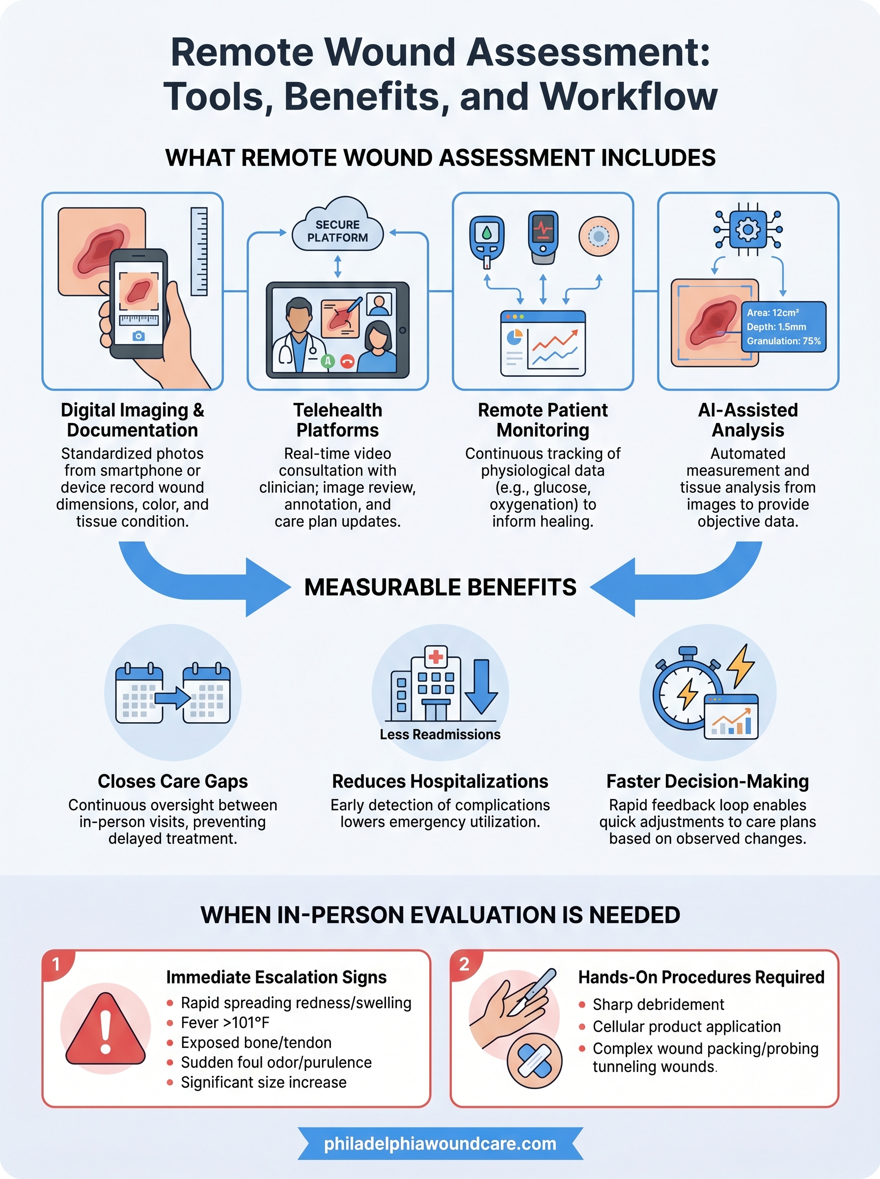
Remote wound assessment is not a single tool or a single conversation with a clinician over a video call. It is a structured clinical process that combines digital imaging, telecommunications platforms, monitoring hardware, and increasingly, AI-assisted analysis software to capture wound data and support clinical decision-making from a distance. Each component serves a distinct function, and when they work together, they give clinicians a picture of wound status that is detailed enough to guide treatment changes, flag complications early, and reduce unnecessary in-person visits.
Digital imaging and photo-based documentation
The foundation of any remote wound assessment is an accurate image. Caregivers, nurses, or patients themselves use a smartphone or dedicated wound imaging device to capture photographs of the wound at scheduled intervals. These images document wound dimensions, tissue color, periwound skin condition, and the presence of exudate or necrotic tissue. The most reliable systems use standardized protocols that include a scale reference, consistent lighting conditions, and specific angles to reduce variability between photos taken on different days.
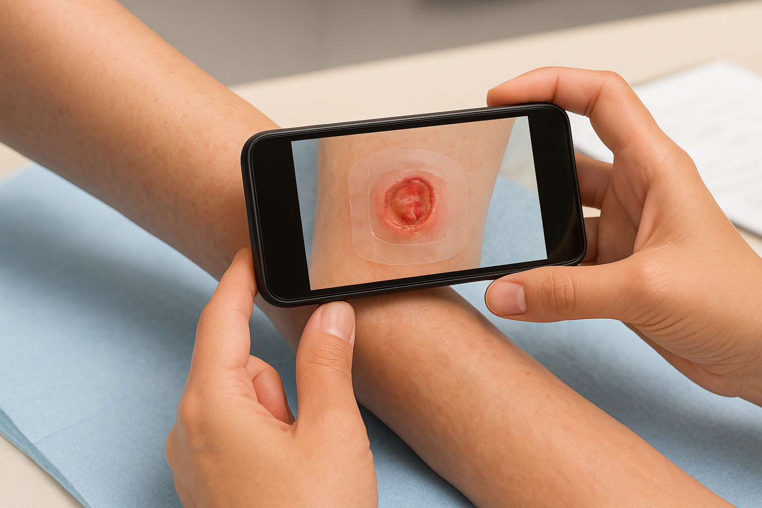
Photo documentation alone gives clinicians a visual record of whether a wound is contracting, granulating, or deteriorating. When images are uploaded to a secure platform with timestamps and patient identifiers, they become a clinical documentation trail that supports billing, care coordination, and any future audits. Facilities and home care teams that skip standardization often end up with photos that are difficult to compare and clinically unreliable.
Consistent photo protocols matter more than the equipment used. A midrange smartphone with a defined process outperforms an advanced camera used inconsistently.
Telehealth platforms and real-time consultation
Secure telehealth platforms allow the clinician and the patient or caregiver to connect in real time via video, review wound images together, and update the care plan without requiring a physical visit. These platforms, when purpose-built for wound care, include tools to annotate images, record measurements, and log clinical findings directly into the patient record. The clinician can ask targeted questions about pain levels, drainage volume, odor, and any recent changes in the surrounding tissue.
Synchronous video visits are most effective when a caregiver or facility nurse is present at the patient’s side during the call. That person becomes the clinician’s hands on the ground, positioning the camera, palpating the wound margins if instructed, and reporting observations the patient may not be able to articulate. The combination of live video and a prepared clinical partner makes the assessment far more accurate than a static image review alone.
Remote patient monitoring systems
Beyond imaging, remote patient monitoring (RPM) systems collect physiological data that informs wound healing indirectly. Devices that track blood glucose levels, peripheral oxygenation, or skin temperature around a wound give clinicians measurable indicators of healing capacity. For diabetic patients, glucose trends captured by continuous monitors are directly relevant to wound healing rates and infection risk.
RPM data streams into a centralized dashboard that the clinical team reviews between scheduled visits. Abnormal readings trigger alerts, allowing the team to contact the patient or caregiver before a problem becomes a hospitalization. Some platforms also capture patient-reported outcomes through short digital questionnaires that ask about pain, wound appearance changes, or dressing performance on a daily or weekly basis.
AI-assisted wound analysis
AI tools now assist clinicians by automatically measuring wound area and depth from uploaded images, flagging tissue types present in a photo, and tracking healing trajectories over time. These tools do not replace clinical judgment, but they reduce the manual measurement burden and add a layer of consistency that human reviewers sometimes miss across a high-volume caseload.
When integrated into a remote wound assessment workflow, AI analysis gives clinicians quantitative data rather than subjective impressions. A wound that looks "about the same" to a caregiver may show a measurable 12% reduction in surface area according to the algorithm, or conversely, a slight increase that warrants closer attention.
Why remote wound assessment matters
Wound healing does not pause between clinic appointments. For patients who are homebound, living in a skilled nursing facility, or in hospice, weeks can pass without a qualified clinician physically reviewing a wound. During that time, infections develop, tissue becomes necrotic, and conditions that were manageable at discharge become emergencies. Remote wound assessment exists to fill that interval with actual clinical oversight rather than hope that nothing changes.
It closes the gap between in-person visits
Most patients with chronic wounds see a clinician once every one to four weeks, depending on their care plan and insurance coverage. That schedule leaves long stretches where caregivers are the only observers. If those caregivers lack the clinical training to recognize early signs of infection or stalled granulation, problems go unreported until the next scheduled visit. Remote assessment tools give your clinical team visibility into the wound on a near-continuous basis, so they can act on changes rather than react to outcomes.
The most dangerous period in wound care is the time between visits when no trained eye is watching.
It reduces preventable hospitalizations
Readmission rates for patients with complex wounds are disproportionately high, and many of those admissions trace back to delayed detection of complications. A wound that receives earlier clinical attention, even through a digital channel, is far less likely to progress to sepsis or require surgical debridement. Studies and payer data consistently show that remote monitoring programs lower emergency utilization for wound care patients, which matters both to patients and to the facilities and family members coordinating their care.
It gives you and your care team faster decision-making data
When your clinician can review a wound photo uploaded at 8 a.m. and call back by noon with updated instructions, the care plan stays current. Without that capability, dressing changes and treatment protocols often continue unchanged past the point where they are still appropriate. Remote assessment compresses the feedback loop between what is observed at the bedside and what the physician orders next. For facilities managing multiple wound care patients across a shift, that speed difference directly affects how many complications get caught before they escalate.
Tools used for remote wound assessment
The clinical categories covered earlier describe what remote wound assessment accomplishes. The tools below are the specific technologies your care team uses in practice. Understanding each one helps you ask sharper questions when evaluating a remote program, and it prepares facility staff and family caregivers to know what to expect when a digital assessment is scheduled for the first time.
Smartphone apps and wound imaging devices
Standard smartphones, when paired with a purpose-built wound care application, can capture color-calibrated images, generate measured overlays, and document tissue type in a format that supports clinical decision-making. These apps differ from a basic camera by attaching objective reference points to each image, so your clinician receives surface area data rather than a subjective visual impression. Some programs also use clip-on structured light attachments that add depth measurement to standard 2D photos.
Your care team should give you specific instructions on which application to use and how to position the wound in frame. Inconsistent technique is the most common reason wound images fail to support an accurate clinical assessment, regardless of how advanced the software is.
Telehealth and remote patient monitoring platforms
HIPAA-compliant video platforms designed for clinical use let your physician review wound images during a live session, annotate findings directly on screen, and update orders before the call ends. General consumer video tools lack the documentation infrastructure to support billing or care coordination at a clinical level. RPM hardware, including Bluetooth-connected glucometers, pulse oximeters, and skin temperature sensors, pairs with these platforms to push physiological data into the patient record automatically between visits.
A platform that connects imaging, monitoring data, and physician notes in a single record is more clinically useful than three separate tools that don’t communicate with each other.
AI-powered wound measurement software
AI measurement tools analyze uploaded images to generate objective data on wound surface area, perimeter, and tissue composition without requiring your clinician to manually trace and calculate each measurement. Clinicians use these outputs to confirm whether a wound is contracting on schedule or stalling. The software also highlights color variations that may signal early infection or inadequate tissue perfusion, giving your care team a reason to intervene before the next scheduled visit rather than discovering the problem after it worsens.
How a remote wound assessment workflow works
A remote wound assessment does not happen spontaneously. It follows a defined sequence that starts before the first image is ever taken and continues until a wound is closed or care transitions to a different setting. Understanding that sequence helps you prepare for what each step requires from your side and what your clinical team does with the information you provide.
Setting up the initial baseline
Before ongoing monitoring begins, your clinician needs a baseline record that documents the wound’s starting condition. This typically happens during an in-person evaluation, whether that is a hospital discharge visit, an initial house call, or a facility intake assessment. The clinician photographs the wound under controlled conditions, measures its dimensions, grades the tissue type present, and records any surrounding skin changes. That baseline becomes the reference point against which every future remote image is compared.
Your care team will also configure the monitoring platform during this phase. That means confirming which devices will collect data, training whoever will capture images at the bedside, and setting the schedule for check-ins. Getting this setup right from the start saves your team from dealing with technically unusable photos or missing data later.
A weak baseline makes every subsequent assessment harder to interpret, so the initial documentation step is worth doing thoroughly.
Ongoing monitoring and clinical response
Once the baseline is in place, the workflow moves into a repeating cycle. At set intervals, your caregiver or facility nurse captures a standardized wound photo using the designated app and uploads it to the secure platform. Physiological monitoring devices push supporting data, such as glucose readings or oxygenation levels, into the same patient record automatically.
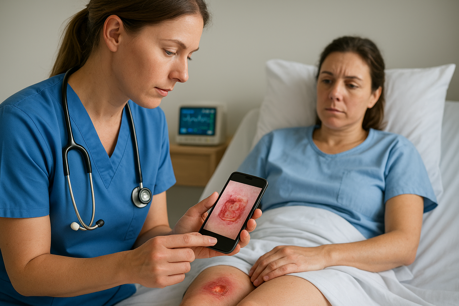
Your clinician reviews the combined data within a defined response window, typically within 24 hours. If the wound is tracking on schedule, the current care plan continues. If the review flags a concern, such as an increase in wound surface area, a color change suggesting infection, or a drop in tissue oxygenation, your clinician contacts your care team directly to update orders, adjust the dressing protocol, or schedule an urgent in-person visit.
This review-and-respond cycle repeats throughout the course of treatment. Documented findings from each cycle feed into the patient record and support billing, reporting to referring physicians, and any transitions between care settings that require a full handoff of wound history.
What clinicians look for in wound photos
A wound photo is only as useful as what a trained clinician can extract from it. During a remote wound assessment, your physician is not just confirming the wound exists; they are pulling specific clinical data from each image that guides the next treatment decision. Knowing what your clinician looks for helps you capture better images and understand why the photo protocol your care team gave you matters.
Tissue type and wound bed condition
The wound bed itself tells your clinician the most important part of the story. Granulation tissue, which appears as pink or red beefy tissue, signals active healing. Yellow slough or black eschar indicates necrotic material that blocks the healing process and may require debridement. Your clinician uses the proportions of these tissue types across the wound bed to gauge whether the current treatment plan is working or needs to change.
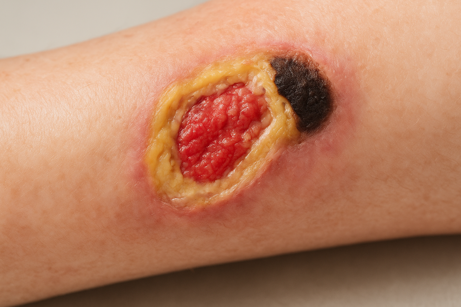
A wound bed shifting from yellow slough toward pink granulation is one of the clearest visual signs that the care plan is moving in the right direction.
Wound edges and surrounding skin
Beyond the wound bed, your clinician examines the wound margins and the skin immediately surrounding the wound. Rolled or undermined wound edges suggest the tissue is not attaching to the wound base, which stalls healing regardless of how well the wound bed looks. Periwound erythema, maceration, or induration points to problems with moisture management, dressing fit, or early infection spreading outward from the wound.
These details require close-up images taken at a consistent angle. Your care team should specify exactly how far to hold the camera from the wound and whether a side-angle shot is needed in addition to the standard overhead view.
Exudate volume and color
Your clinician also evaluates exudate characteristics from what is visible in the photo combined with your reported observations. High-volume drainage or a shift from clear to cloudy or colored exudate changes the clinical picture immediately. Purulent or foul-smelling drainage that a caregiver reports alongside the image gives the physician grounds to escalate care, change the antimicrobial dressing, or schedule an urgent in-person evaluation before the next check-in is due.
Capturing photos before the old dressing is fully removed, and again after the wound is cleaned, gives your clinician the most complete view of what is actively happening at the wound site between visits.
Safety, privacy, and documentation basics
Running a remote wound assessment through digital channels introduces responsibilities that go beyond clinical accuracy. Every image, data point, and clinical note transmitted between a patient’s bedside and a physician must travel through secure, compliant infrastructure. Getting this part right protects the patient, protects your care team, and ensures that the documentation your practice generates holds up during billing reviews or insurance audits.
HIPAA compliance and platform requirements
Any platform your team uses to transmit wound images or patient health information must meet HIPAA requirements under the Health Insurance Portability and Accountability Act, which governs how protected health information is stored, transmitted, and accessed. Consumer video or photo-sharing tools, even widely used ones, do not meet this standard without a signed Business Associate Agreement (BAA) from the vendor. Before your care team sends a single wound image through any digital channel, confirm that the platform your physician uses has that agreement in place.
Using a non-compliant channel to share wound photos, even once, creates a reportable breach that can trigger penalties and erode patient trust.
Your care team should also control who has access to the patient’s wound record within the platform. Access permissions should match clinical roles, so a facility nurse who uploads images does not have the same record access as the supervising physician. Most enterprise-grade clinical platforms handle this through role-based access controls, but confirming the configuration for your specific workflow is your responsibility during setup.
What your documentation record should include
Every remote wound assessment session needs a documented entry in the patient record that captures the date and time of the review, the clinical findings the physician identified, any changes made to the care plan, and who was present or responsible for the bedside observation. This entry should reference the specific images reviewed, not just note that photos were submitted. Linking findings directly to images gives the record clinical substance that supports billing and makes handoffs between providers accurate.
Your documentation should also capture any patient or caregiver reports collected during the session, including pain scores, drainage observations, and dressing performance. These subjective reports become part of the clinical picture and give referring physicians the full context they need to make decisions about next steps in care.
When remote assessment is not enough
Remote wound assessment is a powerful tool for monitoring and early intervention, but it has clinical limits. Digital images and physiological data inform decisions, but they cannot replace what a physician’s hands, nose, and eyes detect at the bedside. Recognizing when to escalate from remote monitoring to direct evaluation protects your patient from preventable deterioration.
Signs that require immediate in-person evaluation
Some wound changes signal a problem serious enough that waiting for the next scheduled remote check-in is not safe. If your caregiver reports any of the following alongside a wound image, your clinical team should treat it as a reason for urgent contact rather than routine review:
- Rapidly spreading redness, warmth, or swelling in the skin surrounding the wound
- Fever above 101°F in a patient with a known wound, combined with increased drainage
- Exposed bone, tendon, or joint tissue visible in the wound bed
- Sudden foul odor or purulent discharge that was not present at the last assessment
- A wound that has increased significantly in size within 24 to 48 hours
When a wound changes fast, digital review should prompt a same-day call, not a note added to the next scheduled session.
These presentations carry infection or tissue loss risk that a photo and a care plan adjustment cannot adequately address. Your clinician needs direct access to the wound to assess depth, probe for undermining, and collect a tissue sample if infection needs to be confirmed.
Wounds that need hands-on intervention
Certain wound conditions require physical procedures that no remote platform can deliver. Sharp debridement of necrotic tissue, application of cellular or tissue-based products under physician supervision, and wound packing adjustments in complex surgical sites all demand that a qualified clinician be present at the patient’s bedside.
Tunneling wounds, those with sinus tracts that extend beyond the visible surface, are particularly difficult to assess remotely because their depth and direction cannot be determined from a photograph alone. A clinician must probe these wounds directly to understand how far they extend and whether the surrounding tissue is undermined. Attempting to manage a tunneling wound through image review alone risks missing a deeper infection or structural involvement that changes the entire treatment approach.
Knowing the boundaries of what remote tools can accomplish is part of using them well. When your care team identifies a wound that exceeds those boundaries, the right response is to get a clinician to the bedside promptly.
Next steps
Remote wound assessment extends clinical oversight into the spaces between visits, but it works best when it connects to a care team that can actually act on what it finds. The tools covered in this article, from AI-powered imaging to RPM dashboards, all depend on a physician with the expertise and access to interpret the data and respond quickly when a wound changes course.
If your patient or loved one has a chronic or non-healing wound and faces barriers getting to a clinic, you do not have to choose between waiting and traveling. Philadelphia Wound Care brings physician-led wound evaluation and advanced treatment directly to homes, skilled nursing facilities, and hospice settings across the Philadelphia region. Our team responds within 24 hours and coordinates directly with referring providers so nothing falls through the gap between visits. To learn more or get started, visit our mobile wound care services in Philadelphia page.